Potassium »
PDB 4zun-5avw »
5a1i »
Potassium in PDB 5a1i: The Structure of Human MAT2A in Complex with Sam, Adenosine, Methionine and Ppnp.
Enzymatic activity of The Structure of Human MAT2A in Complex with Sam, Adenosine, Methionine and Ppnp.
All present enzymatic activity of The Structure of Human MAT2A in Complex with Sam, Adenosine, Methionine and Ppnp.:
2.5.1.6;
2.5.1.6;
Protein crystallography data
The structure of The Structure of Human MAT2A in Complex with Sam, Adenosine, Methionine and Ppnp., PDB code: 5a1i
was solved by
B.Murray,
S.V.Antonyuk,
A.Marina,
S.C.Lu,
J.M.Mato,
S.S.Hasnain,
A.L.Rojas,
with X-Ray Crystallography technique. A brief refinement statistics is given in the table below:
| Resolution Low / High (Å) | 73.37 / 1.09 |
| Space group | I 2 2 2 |
| Cell size a, b, c (Å), α, β, γ (°) | 67.972, 94.074, 117.216, 90.00, 90.00, 90.00 |
| R / Rfree (%) | 10.4 / 12.4 |
Other elements in 5a1i:
The structure of The Structure of Human MAT2A in Complex with Sam, Adenosine, Methionine and Ppnp. also contains other interesting chemical elements:
| Magnesium | (Mg) | 2 atoms |
Potassium Binding Sites:
The binding sites of Potassium atom in the The Structure of Human MAT2A in Complex with Sam, Adenosine, Methionine and Ppnp.
(pdb code 5a1i). This binding sites where shown within
5.0 Angstroms radius around Potassium atom.
In total 2 binding sites of Potassium where determined in the The Structure of Human MAT2A in Complex with Sam, Adenosine, Methionine and Ppnp., PDB code: 5a1i:
Jump to Potassium binding site number: 1; 2;
In total 2 binding sites of Potassium where determined in the The Structure of Human MAT2A in Complex with Sam, Adenosine, Methionine and Ppnp., PDB code: 5a1i:
Jump to Potassium binding site number: 1; 2;
Potassium binding site 1 out of 2 in 5a1i
Go back to
Potassium binding site 1 out
of 2 in the The Structure of Human MAT2A in Complex with Sam, Adenosine, Methionine and Ppnp.
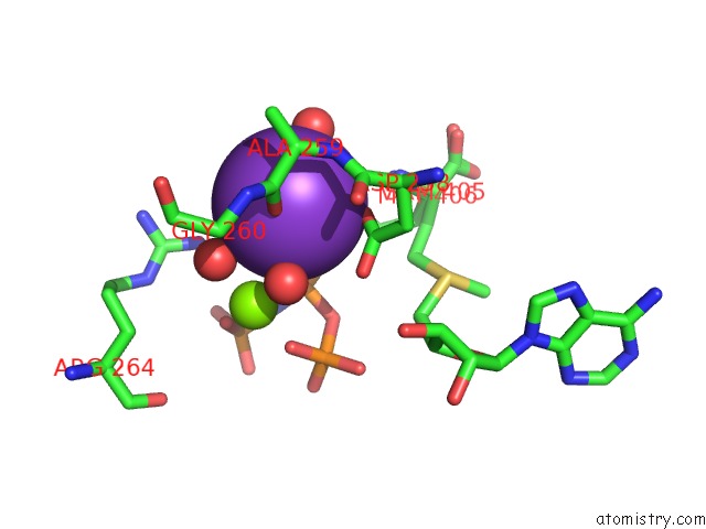
Mono view
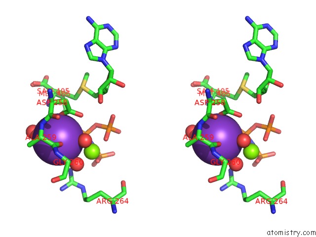
Stereo pair view
Mono view
Stereo pair view
A full contact list of Potassium with other atoms in the K binding
site number 1 of The Structure of Human MAT2A in Complex with Sam, Adenosine, Methionine and Ppnp. within 5.0Å range:
|
Potassium binding site 2 out of 2 in 5a1i
Go back to
Potassium binding site 2 out
of 2 in the The Structure of Human MAT2A in Complex with Sam, Adenosine, Methionine and Ppnp.
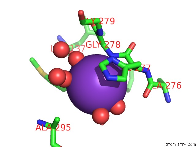
Mono view
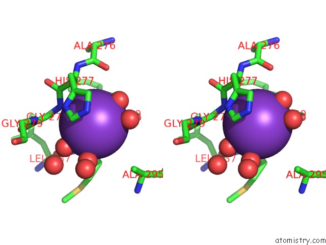
Stereo pair view
Mono view
Stereo pair view
A full contact list of Potassium with other atoms in the K binding
site number 2 of The Structure of Human MAT2A in Complex with Sam, Adenosine, Methionine and Ppnp. within 5.0Å range:
|
Reference:
B.Murray,
S.V.Antonyuk,
A.Marina,
S.C.Lu,
J.M.Mato,
S.S.Hasnain,
A.L.Rojas.
Crystallography Captures Catalytic Steps in Human Methionine Adenosyltransferase Enzymes. Proc.Natl.Acad.Sci.Usa V. 113 2104 2016.
ISSN: ISSN 0027-8424
PubMed: 26858410
DOI: 10.1073/PNAS.1510959113
Page generated: Mon Aug 12 12:51:03 2024
ISSN: ISSN 0027-8424
PubMed: 26858410
DOI: 10.1073/PNAS.1510959113
Last articles
Zn in 9MJ5Zn in 9HNW
Zn in 9G0L
Zn in 9FNE
Zn in 9DZN
Zn in 9E0I
Zn in 9D32
Zn in 9DAK
Zn in 8ZXC
Zn in 8ZUF