Potassium »
PDB 4zun-5avw »
4zxo »
Potassium in PDB 4zxo: The Structure of A GH26 Beta-Mannanase From Bacteroides Ovatus, BOMAN26A.
Protein crystallography data
The structure of The Structure of A GH26 Beta-Mannanase From Bacteroides Ovatus, BOMAN26A., PDB code: 4zxo
was solved by
V.Bagenholm,
O.Aurelius,
D.T.Logan,
H.Bouraoui,
H.Stalbrand,
with X-Ray Crystallography technique. A brief refinement statistics is given in the table below:
| Resolution Low / High (Å) | 41.27 / 1.50 |
| Space group | P 21 21 21 |
| Cell size a, b, c (Å), α, β, γ (°) | 46.856, 79.434, 87.176, 90.00, 90.00, 90.00 |
| R / Rfree (%) | 13.6 / 17.8 |
Potassium Binding Sites:
The binding sites of Potassium atom in the The Structure of A GH26 Beta-Mannanase From Bacteroides Ovatus, BOMAN26A.
(pdb code 4zxo). This binding sites where shown within
5.0 Angstroms radius around Potassium atom.
In total only one binding site of Potassium was determined in the The Structure of A GH26 Beta-Mannanase From Bacteroides Ovatus, BOMAN26A., PDB code: 4zxo:
In total only one binding site of Potassium was determined in the The Structure of A GH26 Beta-Mannanase From Bacteroides Ovatus, BOMAN26A., PDB code: 4zxo:
Potassium binding site 1 out of 1 in 4zxo
Go back to
Potassium binding site 1 out
of 1 in the The Structure of A GH26 Beta-Mannanase From Bacteroides Ovatus, BOMAN26A.
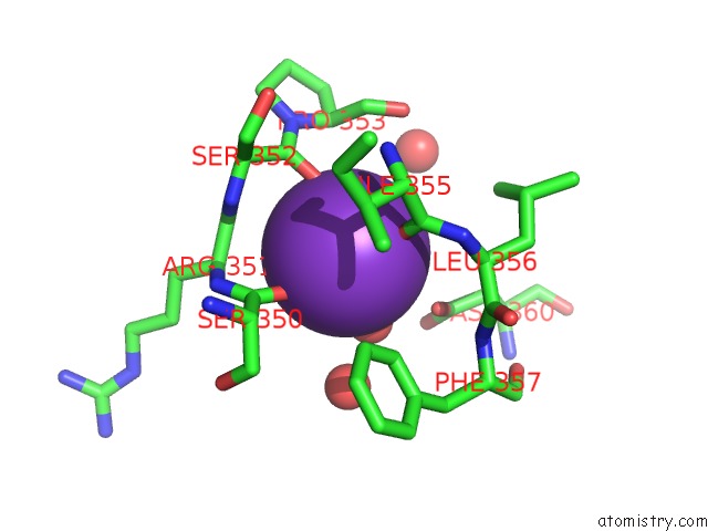
Mono view
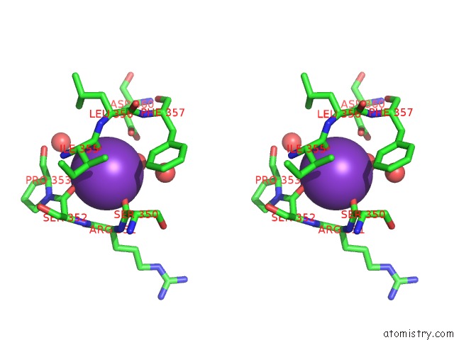
Stereo pair view
Mono view
Stereo pair view
A full contact list of Potassium with other atoms in the K binding
site number 1 of The Structure of A GH26 Beta-Mannanase From Bacteroides Ovatus, BOMAN26A. within 5.0Å range:
|
Reference:
V.Bagenholm,
S.K.Reddy,
H.Bouraoui,
J.Morrill,
E.Kulcinskaja,
C.M.Bahr,
O.Aurelius,
T.Rogers,
Y.Xiao,
D.T.Logan,
E.C.Martens,
N.M.Koropatkin,
H.Stalbrand.
Galactomannan Catabolism Conferred By A Polysaccharide Utilization Locus of Bacteroides Ovatus: Enzyme Synergy and Crystal Structure of A Beta-Mannanase. J. Biol. Chem. V. 292 229 2017.
ISSN: ESSN 1083-351X
PubMed: 27872187
DOI: 10.1074/JBC.M116.746438
Page generated: Mon Aug 12 12:50:38 2024
ISSN: ESSN 1083-351X
PubMed: 27872187
DOI: 10.1074/JBC.M116.746438
Last articles
Zn in 9J0NZn in 9J0O
Zn in 9J0P
Zn in 9FJX
Zn in 9EKB
Zn in 9C0F
Zn in 9CAH
Zn in 9CH0
Zn in 9CH3
Zn in 9CH1