Potassium »
PDB 4xs6-4zum »
4ycl »
Potassium in PDB 4ycl: Crystal Structure of the Sr CA2+-Atpase with Bound Cpa
Enzymatic activity of Crystal Structure of the Sr CA2+-Atpase with Bound Cpa
All present enzymatic activity of Crystal Structure of the Sr CA2+-Atpase with Bound Cpa:
3.6.3.8;
3.6.3.8;
Protein crystallography data
The structure of Crystal Structure of the Sr CA2+-Atpase with Bound Cpa, PDB code: 4ycl
was solved by
H.Ogawa,
M.Takahashi,
Y.Kondou,
C.Toyoshima,
with X-Ray Crystallography technique. A brief refinement statistics is given in the table below:
| Resolution Low / High (Å) | 15.98 / 3.25 |
| Space group | P 1 21 1 |
| Cell size a, b, c (Å), α, β, γ (°) | 63.039, 96.029, 155.418, 90.00, 95.09, 90.00 |
| R / Rfree (%) | 19.7 / 23.7 |
Other elements in 4ycl:
The structure of Crystal Structure of the Sr CA2+-Atpase with Bound Cpa also contains other interesting chemical elements:
| Magnesium | (Mg) | 1 atom |
Potassium Binding Sites:
The binding sites of Potassium atom in the Crystal Structure of the Sr CA2+-Atpase with Bound Cpa
(pdb code 4ycl). This binding sites where shown within
5.0 Angstroms radius around Potassium atom.
In total only one binding site of Potassium was determined in the Crystal Structure of the Sr CA2+-Atpase with Bound Cpa, PDB code: 4ycl:
In total only one binding site of Potassium was determined in the Crystal Structure of the Sr CA2+-Atpase with Bound Cpa, PDB code: 4ycl:
Potassium binding site 1 out of 1 in 4ycl
Go back to
Potassium binding site 1 out
of 1 in the Crystal Structure of the Sr CA2+-Atpase with Bound Cpa
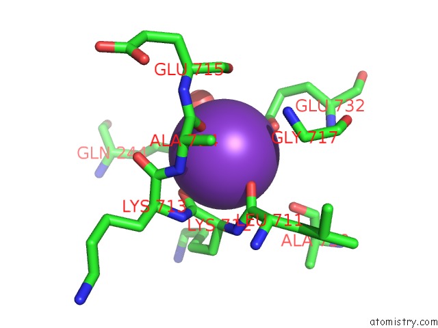
Mono view
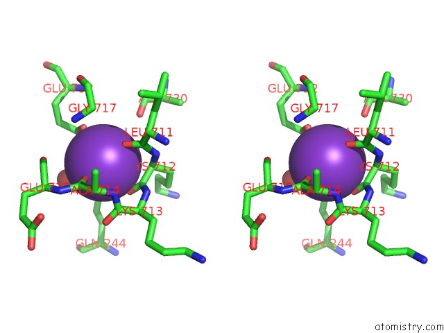
Stereo pair view
Mono view
Stereo pair view
A full contact list of Potassium with other atoms in the K binding
site number 1 of Crystal Structure of the Sr CA2+-Atpase with Bound Cpa within 5.0Å range:
|
Reference:
M.Takahashi,
Y.Kondou,
C.Toyoshima.
Interdomain Communication in Calcium Pump As Revealed in the Crystal Structures with Transmembrane Inhibitors Proc.Natl.Acad.Sci.Usa V. 104 5800 2007.
ISSN: ISSN 0027-8424
PubMed: 17389383
DOI: 10.1073/PNAS.0700979104
Page generated: Sat Aug 9 08:22:02 2025
ISSN: ISSN 0027-8424
PubMed: 17389383
DOI: 10.1073/PNAS.0700979104
Last articles
Mg in 2B2KMg in 2B21
Mg in 2B1J
Mg in 2B1R
Mg in 2B1Q
Mg in 2B0Q
Mg in 2B1D
Mg in 2B1C
Mg in 2B1B
Mg in 2B0T