Potassium »
PDB 4wpz-4xs4 »
4xou »
Potassium in PDB 4xou: Crystal Structure of the Sr CA2+-Atpase in the CA2-E1-Mgamppcp Form Determined By Serial Femtosecond Crystallography Using An X-Ray Free- Electron Laser.
Enzymatic activity of Crystal Structure of the Sr CA2+-Atpase in the CA2-E1-Mgamppcp Form Determined By Serial Femtosecond Crystallography Using An X-Ray Free- Electron Laser.
All present enzymatic activity of Crystal Structure of the Sr CA2+-Atpase in the CA2-E1-Mgamppcp Form Determined By Serial Femtosecond Crystallography Using An X-Ray Free- Electron Laser.:
3.6.3.8;
3.6.3.8;
Protein crystallography data
The structure of Crystal Structure of the Sr CA2+-Atpase in the CA2-E1-Mgamppcp Form Determined By Serial Femtosecond Crystallography Using An X-Ray Free- Electron Laser., PDB code: 4xou
was solved by
M.Bublitz,
K.Nass,
N.D.Drachmann,
A.J.Markvardsen,
M.J.Gutmann,
T.R.M.Barends,
D.Mattle,
R.L.Shoeman,
R.B.Doak,
S.Boutet,
M.Messerschmidt,
M.M.Seibert,
G.J.Williams,
L.Foucar,
L.Reinhard,
O.Sitsel,
J.L.Gregersen,
J.D.Clausen,
T.Boesen,
K.Gotfryd,
K.-T.Wang,
C.Olesen,
J.V.Moller,
P.Nissen,
I.Schlichting,
with X-Ray Crystallography technique. A brief refinement statistics is given in the table below:
| Resolution Low / High (Å) | 59.87 / 2.80 |
| Space group | C 1 2 1 |
| Cell size a, b, c (Å), α, β, γ (°) | 162.000, 76.300, 151.100, 90.00, 109.00, 90.00 |
| R / Rfree (%) | 30.4 / 34.3 |
Other elements in 4xou:
The structure of Crystal Structure of the Sr CA2+-Atpase in the CA2-E1-Mgamppcp Form Determined By Serial Femtosecond Crystallography Using An X-Ray Free- Electron Laser. also contains other interesting chemical elements:
| Calcium | (Ca) | 3 atoms |
Potassium Binding Sites:
The binding sites of Potassium atom in the Crystal Structure of the Sr CA2+-Atpase in the CA2-E1-Mgamppcp Form Determined By Serial Femtosecond Crystallography Using An X-Ray Free- Electron Laser.
(pdb code 4xou). This binding sites where shown within
5.0 Angstroms radius around Potassium atom.
In total only one binding site of Potassium was determined in the Crystal Structure of the Sr CA2+-Atpase in the CA2-E1-Mgamppcp Form Determined By Serial Femtosecond Crystallography Using An X-Ray Free- Electron Laser., PDB code: 4xou:
In total only one binding site of Potassium was determined in the Crystal Structure of the Sr CA2+-Atpase in the CA2-E1-Mgamppcp Form Determined By Serial Femtosecond Crystallography Using An X-Ray Free- Electron Laser., PDB code: 4xou:
Potassium binding site 1 out of 1 in 4xou
Go back to
Potassium binding site 1 out
of 1 in the Crystal Structure of the Sr CA2+-Atpase in the CA2-E1-Mgamppcp Form Determined By Serial Femtosecond Crystallography Using An X-Ray Free- Electron Laser.
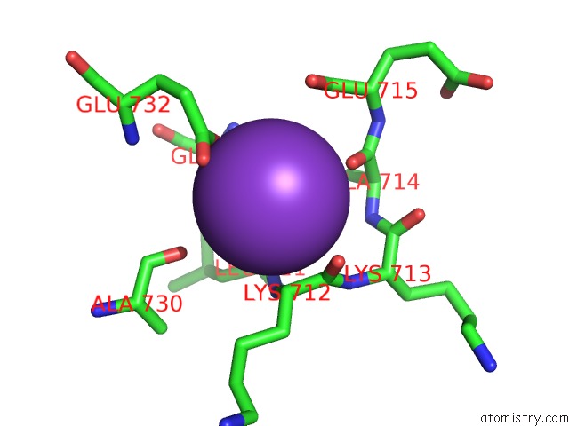
Mono view
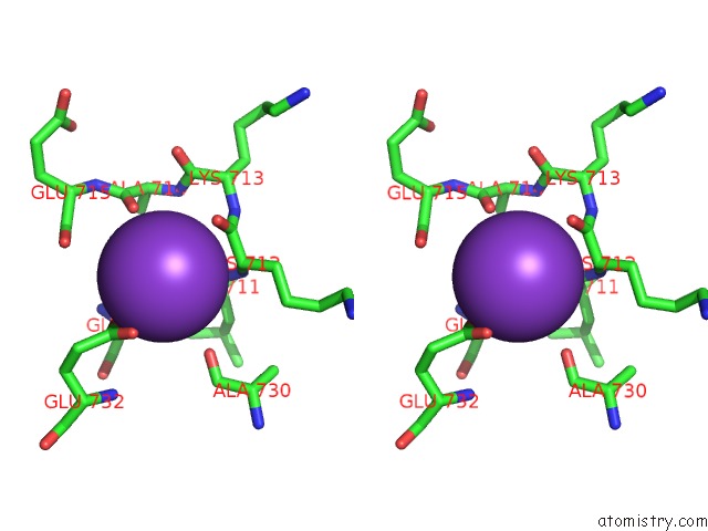
Stereo pair view
Mono view
Stereo pair view
A full contact list of Potassium with other atoms in the K binding
site number 1 of Crystal Structure of the Sr CA2+-Atpase in the CA2-E1-Mgamppcp Form Determined By Serial Femtosecond Crystallography Using An X-Ray Free- Electron Laser. within 5.0Å range:
|
Reference:
M.Bublitz,
K.Nass,
N.D.Drachmann,
A.J.Markvardsen,
M.J.Gutmann,
T.R.Barends,
D.Mattle,
R.L.Shoeman,
R.B.Doak,
S.Boutet,
M.Messerschmidt,
M.M.Seibert,
G.J.Williams,
L.Foucar,
L.Reinhard,
O.Sitsel,
J.L.Gregersen,
J.D.Clausen,
T.Boesen,
K.Gotfryd,
K.T.Wang,
C.Olesen,
J.V.Moller,
P.Nissen,
I.Schlichting.
Structural Studies of P-Type Atpase-Ligand Complexes Using An X-Ray Free-Electron Laser. Iucrj V. 2 409 2015.
ISSN: ESSN 2052-2525
PubMed: 26175901
DOI: 10.1107/S2052252515008969
Page generated: Mon Aug 12 12:36:04 2024
ISSN: ESSN 2052-2525
PubMed: 26175901
DOI: 10.1107/S2052252515008969
Last articles
Zn in 9J0NZn in 9J0O
Zn in 9J0P
Zn in 9FJX
Zn in 9EKB
Zn in 9C0F
Zn in 9CAH
Zn in 9CH0
Zn in 9CH3
Zn in 9CH1