Potassium »
PDB 4toh-4wol »
4uu1 »
Potassium in PDB 4uu1: Crystal Structure of (Sr) Calcium-Atpase E2(Tg) in the Presence of Dopc
Enzymatic activity of Crystal Structure of (Sr) Calcium-Atpase E2(Tg) in the Presence of Dopc
All present enzymatic activity of Crystal Structure of (Sr) Calcium-Atpase E2(Tg) in the Presence of Dopc:
3.6.3.8;
3.6.3.8;
Protein crystallography data
The structure of Crystal Structure of (Sr) Calcium-Atpase E2(Tg) in the Presence of Dopc, PDB code: 4uu1
was solved by
N.D.Drachmann,
C.Olesen,
J.V.Moeller,
Z.Guo,
P.Nissen,
M.Bublitz,
with X-Ray Crystallography technique. A brief refinement statistics is given in the table below:
| Resolution Low / High (Å) | 73.915 / 2.80 |
| Space group | P 41 21 2 |
| Cell size a, b, c (Å), α, β, γ (°) | 71.703, 71.703, 591.318, 90.00, 90.00, 90.00 |
| R / Rfree (%) | 22.11 / 29.29 |
Other elements in 4uu1:
The structure of Crystal Structure of (Sr) Calcium-Atpase E2(Tg) in the Presence of Dopc also contains other interesting chemical elements:
| Magnesium | (Mg) | 1 atom |
Potassium Binding Sites:
The binding sites of Potassium atom in the Crystal Structure of (Sr) Calcium-Atpase E2(Tg) in the Presence of Dopc
(pdb code 4uu1). This binding sites where shown within
5.0 Angstroms radius around Potassium atom.
In total only one binding site of Potassium was determined in the Crystal Structure of (Sr) Calcium-Atpase E2(Tg) in the Presence of Dopc, PDB code: 4uu1:
In total only one binding site of Potassium was determined in the Crystal Structure of (Sr) Calcium-Atpase E2(Tg) in the Presence of Dopc, PDB code: 4uu1:
Potassium binding site 1 out of 1 in 4uu1
Go back to
Potassium binding site 1 out
of 1 in the Crystal Structure of (Sr) Calcium-Atpase E2(Tg) in the Presence of Dopc
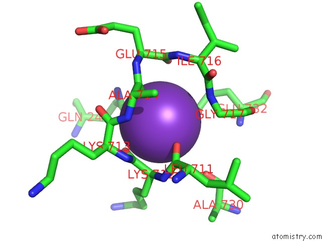
Mono view
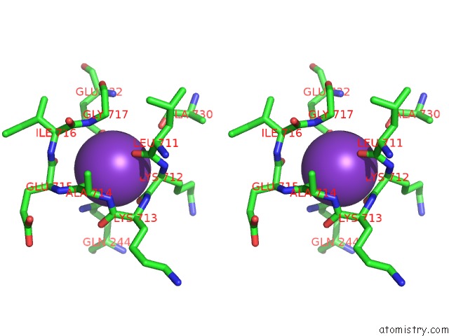
Stereo pair view
Mono view
Stereo pair view
A full contact list of Potassium with other atoms in the K binding
site number 1 of Crystal Structure of (Sr) Calcium-Atpase E2(Tg) in the Presence of Dopc within 5.0Å range:
|
Reference:
N.D.Drachmann,
C.Olesen,
J.V.Moller,
Z.Guo,
P.Nissen,
M.Bublitz.
Comparing Crystal Structures of Ca(2+) -Atpase in the Presence of Different Lipids. Febs J. V. 281 4249 2014.
ISSN: ISSN 1742-464X
PubMed: 25103814
DOI: 10.1111/FEBS.12957
Page generated: Mon Aug 12 12:17:19 2024
ISSN: ISSN 1742-464X
PubMed: 25103814
DOI: 10.1111/FEBS.12957
Last articles
Zn in 9J0NZn in 9J0O
Zn in 9J0P
Zn in 9FJX
Zn in 9EKB
Zn in 9C0F
Zn in 9CAH
Zn in 9CH0
Zn in 9CH3
Zn in 9CH1