Potassium »
PDB 4toh-4wol »
4udi »
Potassium in PDB 4udi: Crystal Structure of B-1,4-Mannopyranosyl-Chitobiose Phosphorylase at 1.85 Angstrom From Unknown Human Gut Bacteria (UHGB_MP)
Protein crystallography data
The structure of Crystal Structure of B-1,4-Mannopyranosyl-Chitobiose Phosphorylase at 1.85 Angstrom From Unknown Human Gut Bacteria (UHGB_MP), PDB code: 4udi
was solved by
S.Ladeveze,
G.Cioci,
G.Potocki-Veronese,
S.Tranier,
L.Mourey,
with X-Ray Crystallography technique. A brief refinement statistics is given in the table below:
| Resolution Low / High (Å) | 110.20 / 1.80 |
| Space group | P 21 21 21 |
| Cell size a, b, c (Å), α, β, γ (°) | 84.116, 141.206, 176.245, 90.00, 90.00, 90.00 |
| R / Rfree (%) | 15.537 / 18.993 |
Potassium Binding Sites:
The binding sites of Potassium atom in the Crystal Structure of B-1,4-Mannopyranosyl-Chitobiose Phosphorylase at 1.85 Angstrom From Unknown Human Gut Bacteria (UHGB_MP)
(pdb code 4udi). This binding sites where shown within
5.0 Angstroms radius around Potassium atom.
In total 6 binding sites of Potassium where determined in the Crystal Structure of B-1,4-Mannopyranosyl-Chitobiose Phosphorylase at 1.85 Angstrom From Unknown Human Gut Bacteria (UHGB_MP), PDB code: 4udi:
Jump to Potassium binding site number: 1; 2; 3; 4; 5; 6;
In total 6 binding sites of Potassium where determined in the Crystal Structure of B-1,4-Mannopyranosyl-Chitobiose Phosphorylase at 1.85 Angstrom From Unknown Human Gut Bacteria (UHGB_MP), PDB code: 4udi:
Jump to Potassium binding site number: 1; 2; 3; 4; 5; 6;
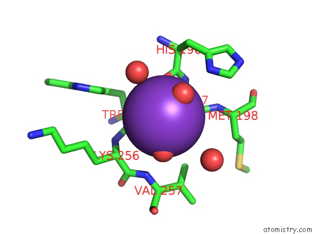
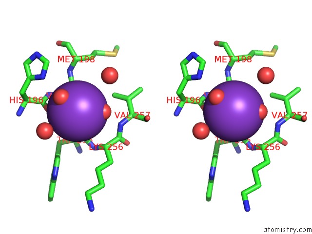
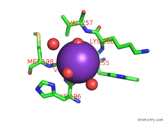
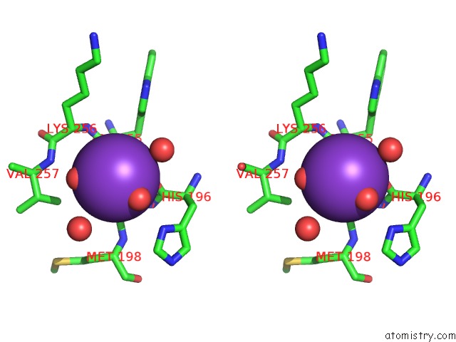
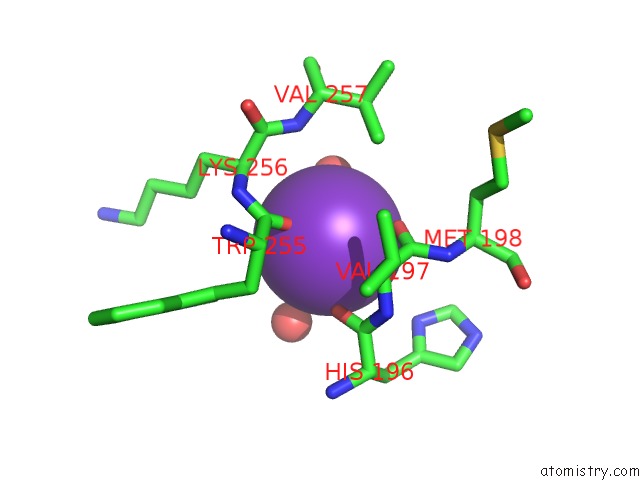
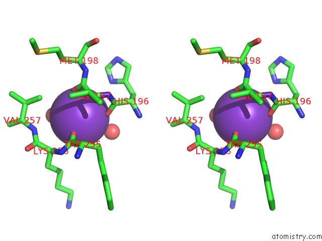
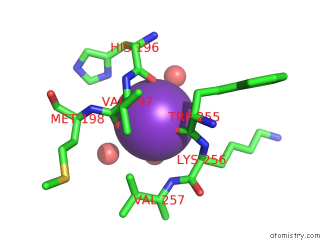
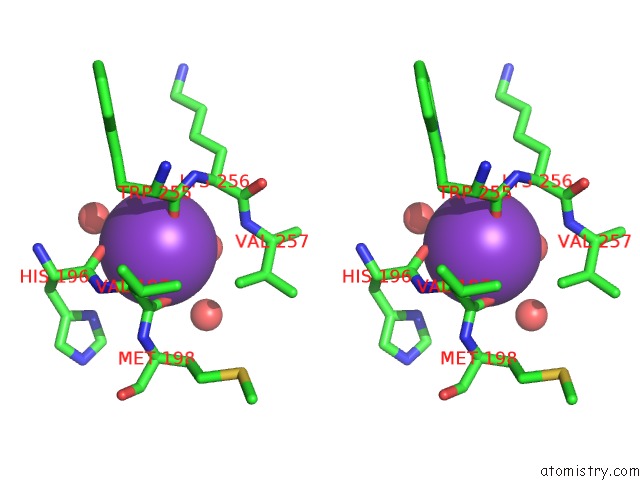
Potassium binding site 1 out of 6 in 4udi
Go back to
Potassium binding site 1 out
of 6 in the Crystal Structure of B-1,4-Mannopyranosyl-Chitobiose Phosphorylase at 1.85 Angstrom From Unknown Human Gut Bacteria (UHGB_MP)
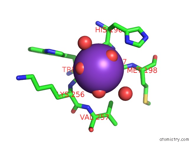
Mono view
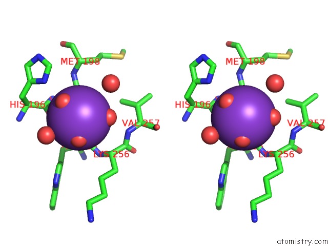
Stereo pair view
Mono view
Stereo pair view
A full contact list of Potassium with other atoms in the K binding
site number 1 of Crystal Structure of B-1,4-Mannopyranosyl-Chitobiose Phosphorylase at 1.85 Angstrom From Unknown Human Gut Bacteria (UHGB_MP) within 5.0Å range:
|
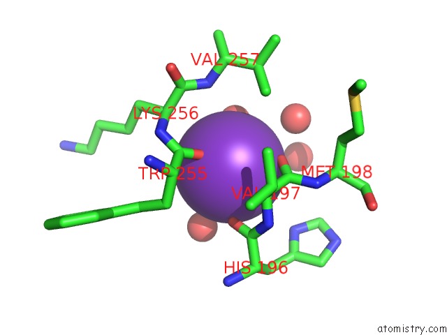
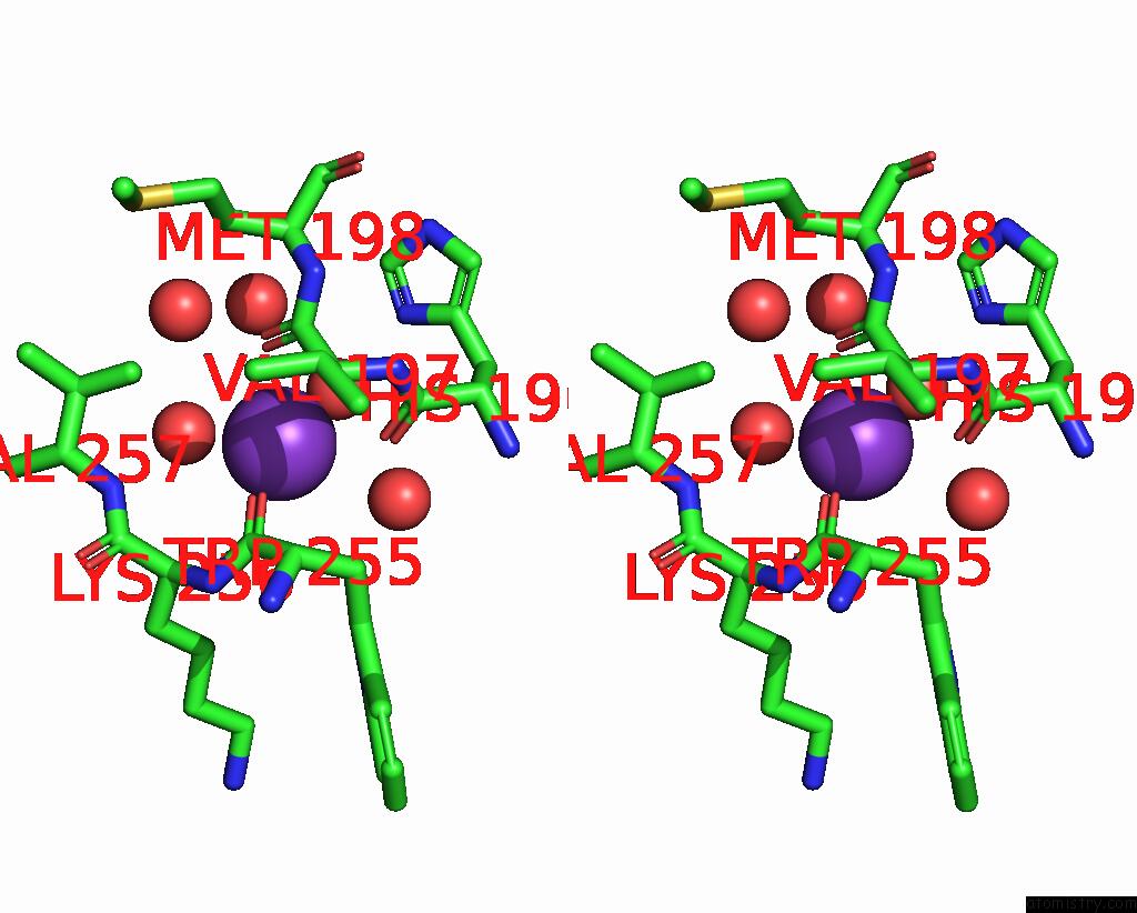
Potassium binding site 2 out of 6 in 4udi
Go back to
Potassium binding site 2 out
of 6 in the Crystal Structure of B-1,4-Mannopyranosyl-Chitobiose Phosphorylase at 1.85 Angstrom From Unknown Human Gut Bacteria (UHGB_MP)
Mono view
Stereo pair view
Mono view
Stereo pair view
A full contact list of Potassium with other atoms in the K binding
site number 2 of Crystal Structure of B-1,4-Mannopyranosyl-Chitobiose Phosphorylase at 1.85 Angstrom From Unknown Human Gut Bacteria (UHGB_MP) within 5.0Å range:
|
Potassium binding site 3 out of 6 in 4udi
Go back to
Potassium binding site 3 out
of 6 in the Crystal Structure of B-1,4-Mannopyranosyl-Chitobiose Phosphorylase at 1.85 Angstrom From Unknown Human Gut Bacteria (UHGB_MP)
Mono view
Stereo pair view
Mono view
Stereo pair view
A full contact list of Potassium with other atoms in the K binding
site number 3 of Crystal Structure of B-1,4-Mannopyranosyl-Chitobiose Phosphorylase at 1.85 Angstrom From Unknown Human Gut Bacteria (UHGB_MP) within 5.0Å range:
|
Potassium binding site 4 out of 6 in 4udi
Go back to
Potassium binding site 4 out
of 6 in the Crystal Structure of B-1,4-Mannopyranosyl-Chitobiose Phosphorylase at 1.85 Angstrom From Unknown Human Gut Bacteria (UHGB_MP)
Mono view
Stereo pair view
Mono view
Stereo pair view
A full contact list of Potassium with other atoms in the K binding
site number 4 of Crystal Structure of B-1,4-Mannopyranosyl-Chitobiose Phosphorylase at 1.85 Angstrom From Unknown Human Gut Bacteria (UHGB_MP) within 5.0Å range:
|
Potassium binding site 5 out of 6 in 4udi
Go back to
Potassium binding site 5 out
of 6 in the Crystal Structure of B-1,4-Mannopyranosyl-Chitobiose Phosphorylase at 1.85 Angstrom From Unknown Human Gut Bacteria (UHGB_MP)
Mono view
Stereo pair view
Mono view
Stereo pair view
A full contact list of Potassium with other atoms in the K binding
site number 5 of Crystal Structure of B-1,4-Mannopyranosyl-Chitobiose Phosphorylase at 1.85 Angstrom From Unknown Human Gut Bacteria (UHGB_MP) within 5.0Å range:
|
Potassium binding site 6 out of 6 in 4udi
Go back to
Potassium binding site 6 out
of 6 in the Crystal Structure of B-1,4-Mannopyranosyl-Chitobiose Phosphorylase at 1.85 Angstrom From Unknown Human Gut Bacteria (UHGB_MP)
Mono view
Stereo pair view
Mono view
Stereo pair view
A full contact list of Potassium with other atoms in the K binding
site number 6 of Crystal Structure of B-1,4-Mannopyranosyl-Chitobiose Phosphorylase at 1.85 Angstrom From Unknown Human Gut Bacteria (UHGB_MP) within 5.0Å range:
|
Reference:
S.Ladeveze,
G.Cioci,
P.Roblin,
L.Mourey,
S.Tranier,
G.Potocki-Veronese.
Structural Bases For N-Glycan Processing By Mannoside Phosphorylase Acta Crystallogr.,Sect.D V. 71 1 2015.
ISSN: ISSN 0907-4449
DOI: 10.1107/S1399004715006604
Page generated: Mon Aug 12 12:14:35 2024
ISSN: ISSN 0907-4449
DOI: 10.1107/S1399004715006604
Last articles
Zn in 9J0NZn in 9J0O
Zn in 9J0P
Zn in 9FJX
Zn in 9EKB
Zn in 9C0F
Zn in 9CAH
Zn in 9CH0
Zn in 9CH3
Zn in 9CH1