Potassium »
PDB 4toh-4wol »
4u5m »
Potassium in PDB 4u5m: Structure of A Left-Handed Dna G-Quadruplex
Protein crystallography data
The structure of Structure of A Left-Handed Dna G-Quadruplex, PDB code: 4u5m
was solved by
E.Schmitt,
Y.Mechulam,
A.T.Phan,
H.Brahim,
W.J.Chung,
K.W.Lim,
with X-Ray Crystallography technique. A brief refinement statistics is given in the table below:
| Resolution Low / High (Å) | 44.94 / 1.50 |
| Space group | P 32 2 1 |
| Cell size a, b, c (Å), α, β, γ (°) | 51.886, 51.886, 40.167, 90.00, 90.00, 120.00 |
| R / Rfree (%) | 14.1 / 18 |
Other elements in 4u5m:
The structure of Structure of A Left-Handed Dna G-Quadruplex also contains other interesting chemical elements:
| Magnesium | (Mg) | 1 atom |
Potassium Binding Sites:
The binding sites of Potassium atom in the Structure of A Left-Handed Dna G-Quadruplex
(pdb code 4u5m). This binding sites where shown within
5.0 Angstroms radius around Potassium atom.
In total 4 binding sites of Potassium where determined in the Structure of A Left-Handed Dna G-Quadruplex, PDB code: 4u5m:
Jump to Potassium binding site number: 1; 2; 3; 4;
In total 4 binding sites of Potassium where determined in the Structure of A Left-Handed Dna G-Quadruplex, PDB code: 4u5m:
Jump to Potassium binding site number: 1; 2; 3; 4;
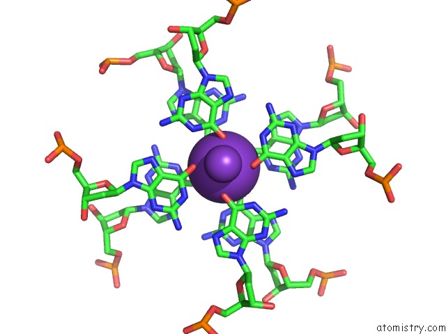
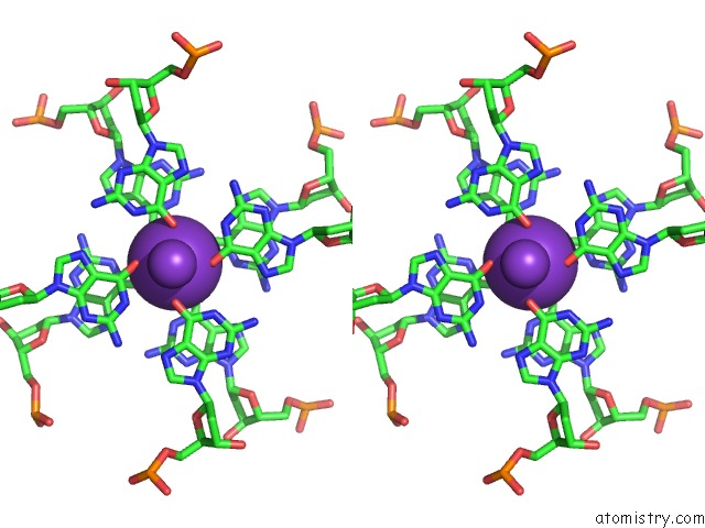
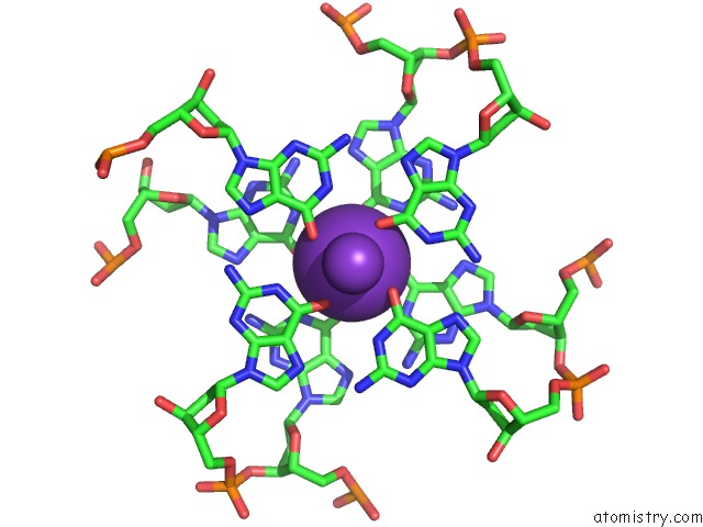
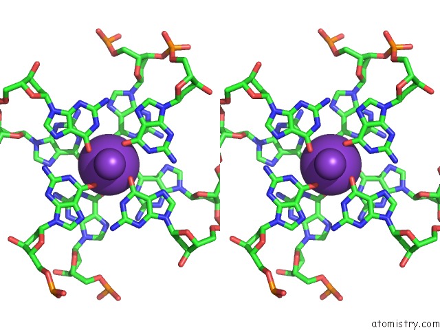
Potassium binding site 1 out of 4 in 4u5m
Go back to
Potassium binding site 1 out
of 4 in the Structure of A Left-Handed Dna G-Quadruplex
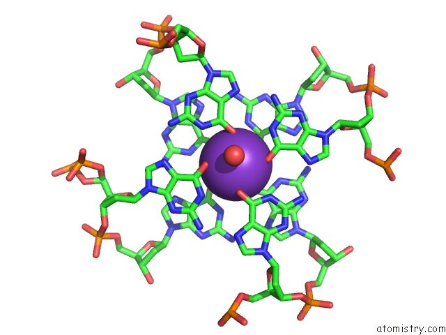
Mono view
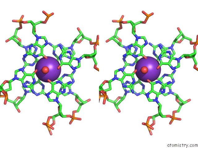
Stereo pair view
Mono view
Stereo pair view
A full contact list of Potassium with other atoms in the K binding
site number 1 of Structure of A Left-Handed Dna G-Quadruplex within 5.0Å range:
|
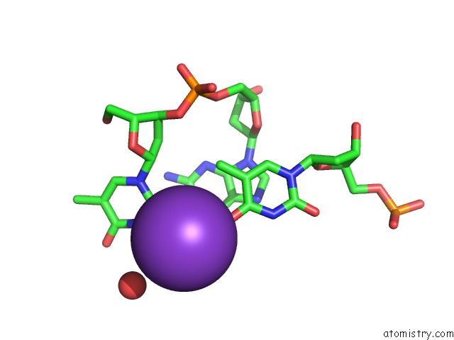
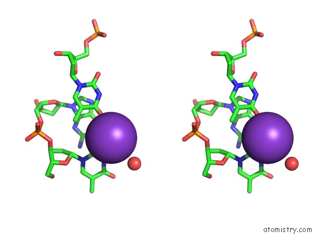
Potassium binding site 2 out of 4 in 4u5m
Go back to
Potassium binding site 2 out
of 4 in the Structure of A Left-Handed Dna G-Quadruplex
Mono view
Stereo pair view
Mono view
Stereo pair view
A full contact list of Potassium with other atoms in the K binding
site number 2 of Structure of A Left-Handed Dna G-Quadruplex within 5.0Å range:
|
Potassium binding site 3 out of 4 in 4u5m
Go back to
Potassium binding site 3 out
of 4 in the Structure of A Left-Handed Dna G-Quadruplex
Mono view
Stereo pair view
Mono view
Stereo pair view
A full contact list of Potassium with other atoms in the K binding
site number 3 of Structure of A Left-Handed Dna G-Quadruplex within 5.0Å range:
|
Potassium binding site 4 out of 4 in 4u5m
Go back to
Potassium binding site 4 out
of 4 in the Structure of A Left-Handed Dna G-Quadruplex
Mono view
Stereo pair view
Mono view
Stereo pair view
A full contact list of Potassium with other atoms in the K binding
site number 4 of Structure of A Left-Handed Dna G-Quadruplex within 5.0Å range:
|
Reference:
W.J.Chung,
B.Heddi,
E.Schmitt,
K.W.Lim,
Y.Mechulam,
A.T.Phan.
Structure of A Left-Handed Dna G-Quadruplex. Proc.Natl.Acad.Sci.Usa 2015.
ISSN: ESSN 1091-6490
PubMed: 25695967
DOI: 10.1073/PNAS.1418718112
Page generated: Mon Aug 12 12:11:23 2024
ISSN: ESSN 1091-6490
PubMed: 25695967
DOI: 10.1073/PNAS.1418718112
Last articles
Zn in 9MJ5Zn in 9HNW
Zn in 9G0L
Zn in 9FNE
Zn in 9DZN
Zn in 9E0I
Zn in 9D32
Zn in 9DAK
Zn in 8ZXC
Zn in 8ZUF