Potassium »
PDB 4qxg-4tog »
4rgf »
Potassium in PDB 4rgf: Crystal Structure of the in-Line Aligned ENV22 Twister Ribozyme Soaked with MN2+
Protein crystallography data
The structure of Crystal Structure of the in-Line Aligned ENV22 Twister Ribozyme Soaked with MN2+, PDB code: 4rgf
was solved by
A.Ren,
K.R.Rajashankar,
D.Simanshu,
D.Patel,
with X-Ray Crystallography technique. A brief refinement statistics is given in the table below:
| Resolution Low / High (Å) | 48.75 / 3.20 |
| Space group | P 61 2 2 |
| Cell size a, b, c (Å), α, β, γ (°) | 64.299, 64.299, 584.991, 90.00, 90.00, 120.00 |
| R / Rfree (%) | 23.1 / 27.7 |
Other elements in 4rgf:
The structure of Crystal Structure of the in-Line Aligned ENV22 Twister Ribozyme Soaked with MN2+ also contains other interesting chemical elements:
| Magnesium | (Mg) | 1 atom |
| Manganese | (Mn) | 11 atoms |
Potassium Binding Sites:
Pages:
>>> Page 1 <<< Page 2, Binding sites: 11 - 14;Binding sites:
The binding sites of Potassium atom in the Crystal Structure of the in-Line Aligned ENV22 Twister Ribozyme Soaked with MN2+ (pdb code 4rgf). This binding sites where shown within 5.0 Angstroms radius around Potassium atom.In total 14 binding sites of Potassium where determined in the Crystal Structure of the in-Line Aligned ENV22 Twister Ribozyme Soaked with MN2+, PDB code: 4rgf:
Jump to Potassium binding site number: 1; 2; 3; 4; 5; 6; 7; 8; 9; 10;
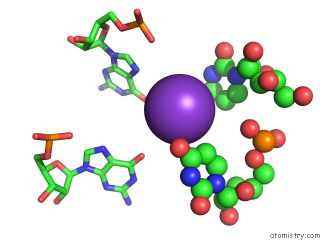
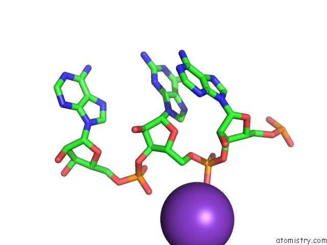
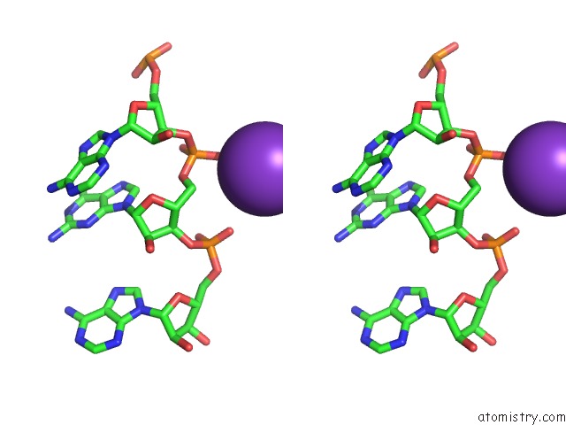
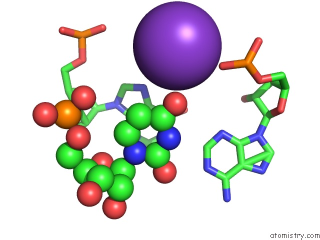
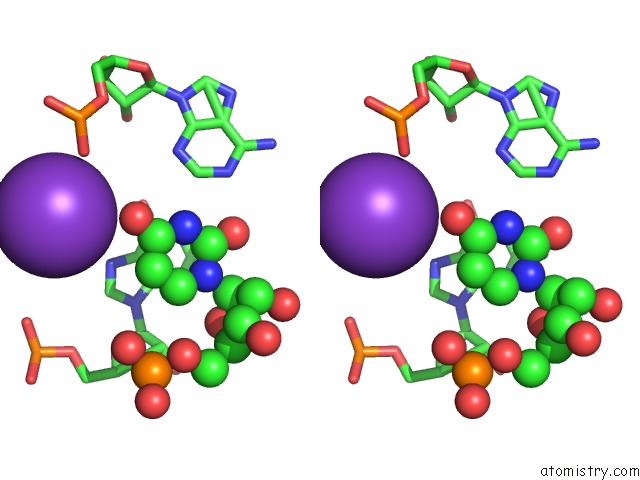
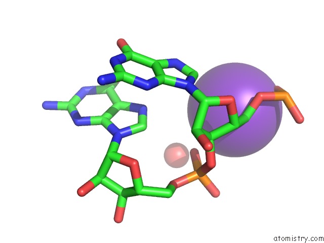
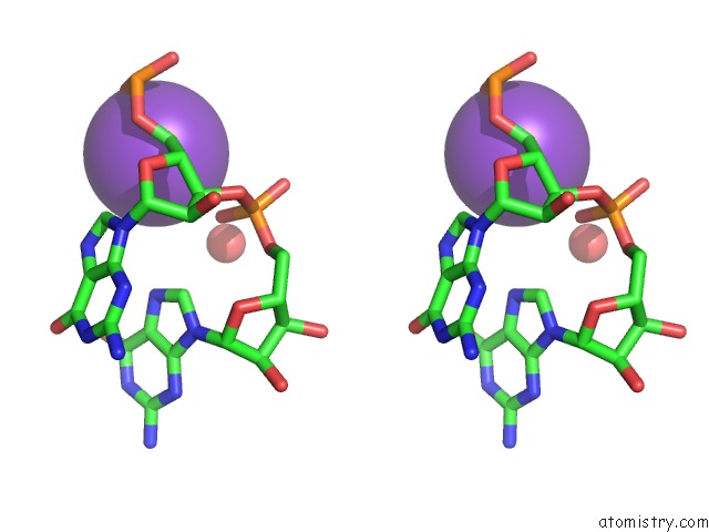
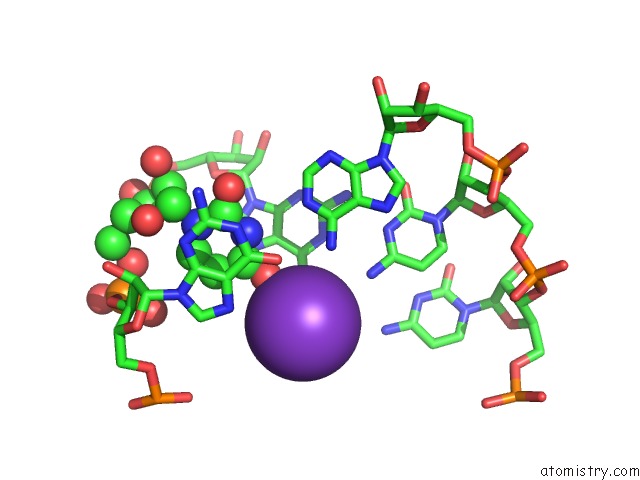
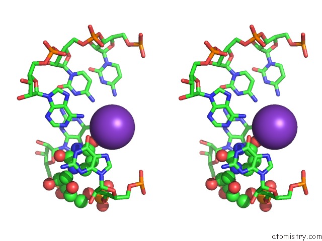
Potassium binding site 1 out of 14 in 4rgf
Go back to
Potassium binding site 1 out
of 14 in the Crystal Structure of the in-Line Aligned ENV22 Twister Ribozyme Soaked with MN2+
Mono view
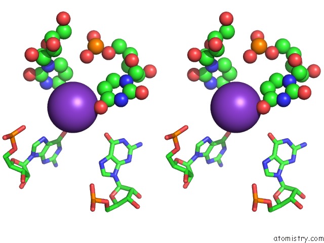
Stereo pair view
Mono view
Stereo pair view
A full contact list of Potassium with other atoms in the K binding
site number 1 of Crystal Structure of the in-Line Aligned ENV22 Twister Ribozyme Soaked with MN2+ within 5.0Å range:
|
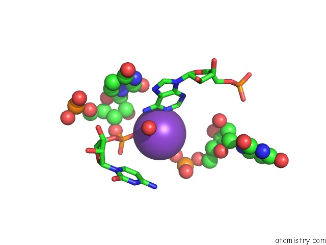
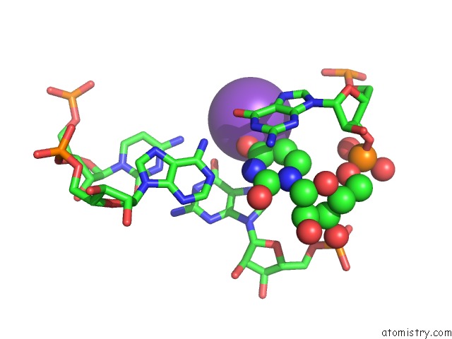
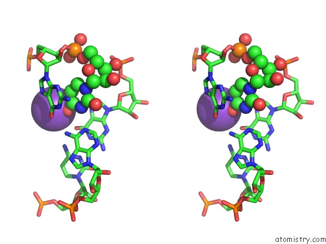
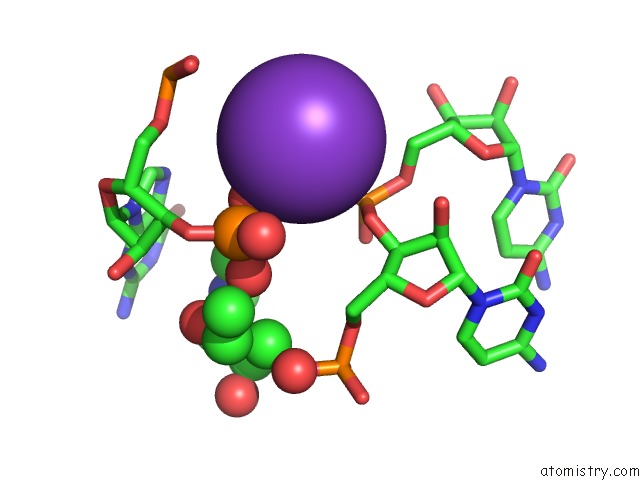
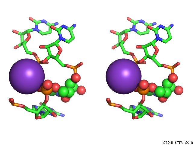
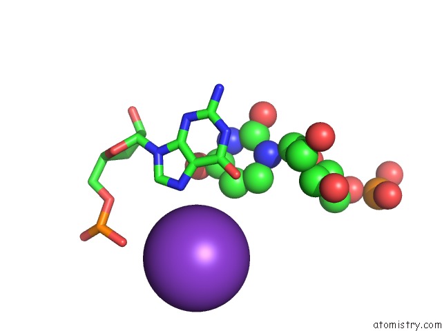
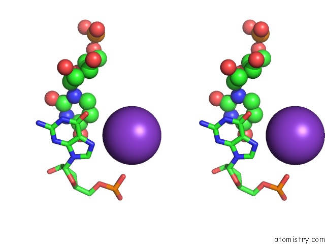
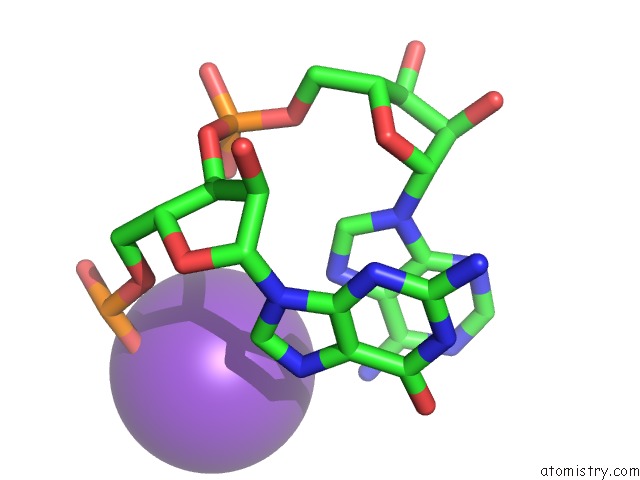
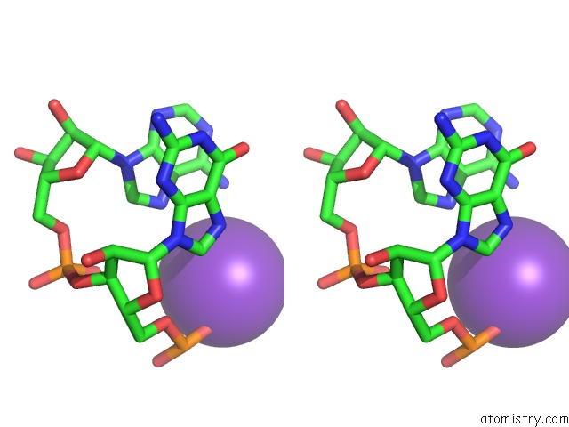
Potassium binding site 2 out of 14 in 4rgf
Go back to
Potassium binding site 2 out
of 14 in the Crystal Structure of the in-Line Aligned ENV22 Twister Ribozyme Soaked with MN2+
Mono view
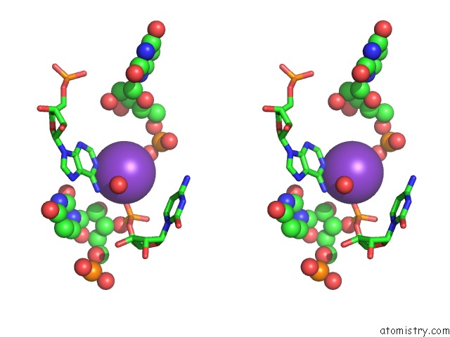
Stereo pair view
Mono view
Stereo pair view
A full contact list of Potassium with other atoms in the K binding
site number 2 of Crystal Structure of the in-Line Aligned ENV22 Twister Ribozyme Soaked with MN2+ within 5.0Å range:
|
Potassium binding site 3 out of 14 in 4rgf
Go back to
Potassium binding site 3 out
of 14 in the Crystal Structure of the in-Line Aligned ENV22 Twister Ribozyme Soaked with MN2+
Mono view
Stereo pair view
Mono view
Stereo pair view
A full contact list of Potassium with other atoms in the K binding
site number 3 of Crystal Structure of the in-Line Aligned ENV22 Twister Ribozyme Soaked with MN2+ within 5.0Å range:
|
Potassium binding site 4 out of 14 in 4rgf
Go back to
Potassium binding site 4 out
of 14 in the Crystal Structure of the in-Line Aligned ENV22 Twister Ribozyme Soaked with MN2+
Mono view
Stereo pair view
Mono view
Stereo pair view
A full contact list of Potassium with other atoms in the K binding
site number 4 of Crystal Structure of the in-Line Aligned ENV22 Twister Ribozyme Soaked with MN2+ within 5.0Å range:
|
Potassium binding site 5 out of 14 in 4rgf
Go back to
Potassium binding site 5 out
of 14 in the Crystal Structure of the in-Line Aligned ENV22 Twister Ribozyme Soaked with MN2+
Mono view
Stereo pair view
Mono view
Stereo pair view
A full contact list of Potassium with other atoms in the K binding
site number 5 of Crystal Structure of the in-Line Aligned ENV22 Twister Ribozyme Soaked with MN2+ within 5.0Å range:
|
Potassium binding site 6 out of 14 in 4rgf
Go back to
Potassium binding site 6 out
of 14 in the Crystal Structure of the in-Line Aligned ENV22 Twister Ribozyme Soaked with MN2+
Mono view
Stereo pair view
Mono view
Stereo pair view
A full contact list of Potassium with other atoms in the K binding
site number 6 of Crystal Structure of the in-Line Aligned ENV22 Twister Ribozyme Soaked with MN2+ within 5.0Å range:
|
Potassium binding site 7 out of 14 in 4rgf
Go back to
Potassium binding site 7 out
of 14 in the Crystal Structure of the in-Line Aligned ENV22 Twister Ribozyme Soaked with MN2+
Mono view
Stereo pair view
Mono view
Stereo pair view
A full contact list of Potassium with other atoms in the K binding
site number 7 of Crystal Structure of the in-Line Aligned ENV22 Twister Ribozyme Soaked with MN2+ within 5.0Å range:
|
Potassium binding site 8 out of 14 in 4rgf
Go back to
Potassium binding site 8 out
of 14 in the Crystal Structure of the in-Line Aligned ENV22 Twister Ribozyme Soaked with MN2+
Mono view
Stereo pair view
Mono view
Stereo pair view
A full contact list of Potassium with other atoms in the K binding
site number 8 of Crystal Structure of the in-Line Aligned ENV22 Twister Ribozyme Soaked with MN2+ within 5.0Å range:
|
Potassium binding site 9 out of 14 in 4rgf
Go back to
Potassium binding site 9 out
of 14 in the Crystal Structure of the in-Line Aligned ENV22 Twister Ribozyme Soaked with MN2+
Mono view
Stereo pair view
Mono view
Stereo pair view
A full contact list of Potassium with other atoms in the K binding
site number 9 of Crystal Structure of the in-Line Aligned ENV22 Twister Ribozyme Soaked with MN2+ within 5.0Å range:
|
Potassium binding site 10 out of 14 in 4rgf
Go back to
Potassium binding site 10 out
of 14 in the Crystal Structure of the in-Line Aligned ENV22 Twister Ribozyme Soaked with MN2+
Mono view
Stereo pair view
Mono view
Stereo pair view
A full contact list of Potassium with other atoms in the K binding
site number 10 of Crystal Structure of the in-Line Aligned ENV22 Twister Ribozyme Soaked with MN2+ within 5.0Å range:
|
Reference:
A.Ren,
M.Kosutic,
K.R.Rajashankar,
M.Frener,
T.Santner,
E.Westhof,
R.Micura,
D.J.Patel.
In-Line Alignment and Mg(2+) Coordination at the Cleavage Site of the ENV22 Twister Ribozyme. Nat Commun V. 5 5534 2014.
ISSN: ESSN 2041-1723
PubMed: 25410397
DOI: 10.1038/NCOMMS6534
Page generated: Mon Aug 12 11:59:06 2024
ISSN: ESSN 2041-1723
PubMed: 25410397
DOI: 10.1038/NCOMMS6534
Last articles
Zn in 9MJ5Zn in 9HNW
Zn in 9G0L
Zn in 9FNE
Zn in 9DZN
Zn in 9E0I
Zn in 9D32
Zn in 9DAK
Zn in 8ZXC
Zn in 8ZUF