Potassium »
PDB 4qxg-4tog »
4rfl »
Potassium in PDB 4rfl: Crystal Structure of G1PDH with Nadph From Methanocaldococcus Jannaschii
Enzymatic activity of Crystal Structure of G1PDH with Nadph From Methanocaldococcus Jannaschii
All present enzymatic activity of Crystal Structure of G1PDH with Nadph From Methanocaldococcus Jannaschii:
1.1.1.261;
1.1.1.261;
Protein crystallography data
The structure of Crystal Structure of G1PDH with Nadph From Methanocaldococcus Jannaschii, PDB code: 4rfl
was solved by
V.Carbone,
R.S.Ronimus,
L.R.Schofield,
A.J.Sutherland-Smith,
with X-Ray Crystallography technique. A brief refinement statistics is given in the table below:
| Resolution Low / High (Å) | 46.68 / 2.20 |
| Space group | P 1 |
| Cell size a, b, c (Å), α, β, γ (°) | 59.391, 72.115, 101.714, 77.62, 79.58, 75.63 |
| R / Rfree (%) | 18.5 / 22.5 |
Other elements in 4rfl:
The structure of Crystal Structure of G1PDH with Nadph From Methanocaldococcus Jannaschii also contains other interesting chemical elements:
| Zinc | (Zn) | 4 atoms |
| Sodium | (Na) | 1 atom |
Potassium Binding Sites:
The binding sites of Potassium atom in the Crystal Structure of G1PDH with Nadph From Methanocaldococcus Jannaschii
(pdb code 4rfl). This binding sites where shown within
5.0 Angstroms radius around Potassium atom.
In total 4 binding sites of Potassium where determined in the Crystal Structure of G1PDH with Nadph From Methanocaldococcus Jannaschii, PDB code: 4rfl:
Jump to Potassium binding site number: 1; 2; 3; 4;
In total 4 binding sites of Potassium where determined in the Crystal Structure of G1PDH with Nadph From Methanocaldococcus Jannaschii, PDB code: 4rfl:
Jump to Potassium binding site number: 1; 2; 3; 4;
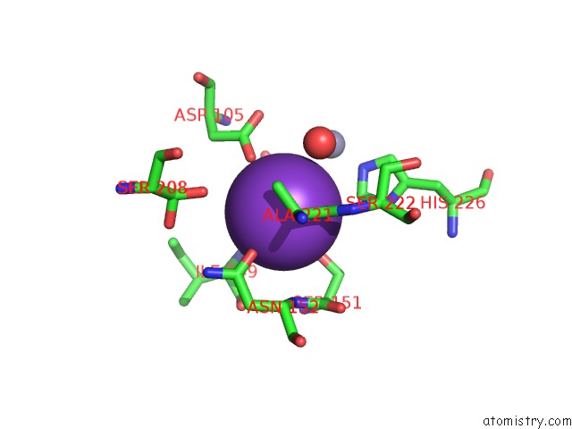
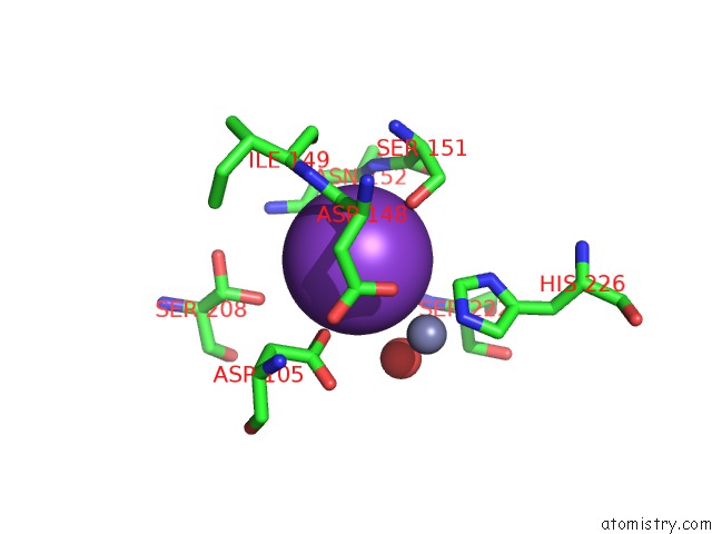
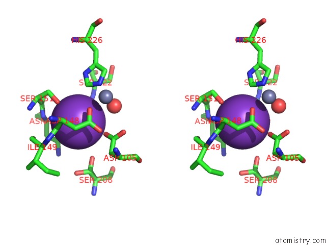
Potassium binding site 1 out of 4 in 4rfl
Go back to
Potassium binding site 1 out
of 4 in the Crystal Structure of G1PDH with Nadph From Methanocaldococcus Jannaschii
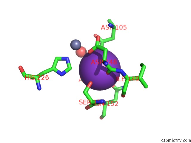
Mono view
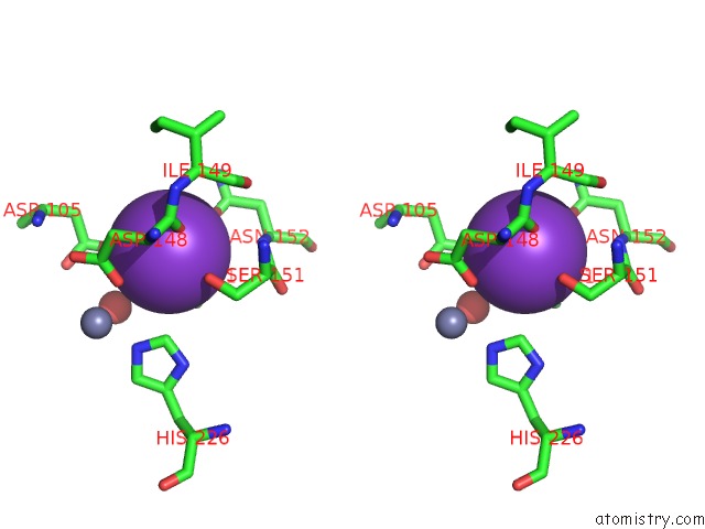
Stereo pair view
Mono view
Stereo pair view
A full contact list of Potassium with other atoms in the K binding
site number 1 of Crystal Structure of G1PDH with Nadph From Methanocaldococcus Jannaschii within 5.0Å range:
|
Potassium binding site 2 out of 4 in 4rfl
Go back to
Potassium binding site 2 out
of 4 in the Crystal Structure of G1PDH with Nadph From Methanocaldococcus Jannaschii
Mono view
Stereo pair view
Mono view
Stereo pair view
A full contact list of Potassium with other atoms in the K binding
site number 2 of Crystal Structure of G1PDH with Nadph From Methanocaldococcus Jannaschii within 5.0Å range:
|
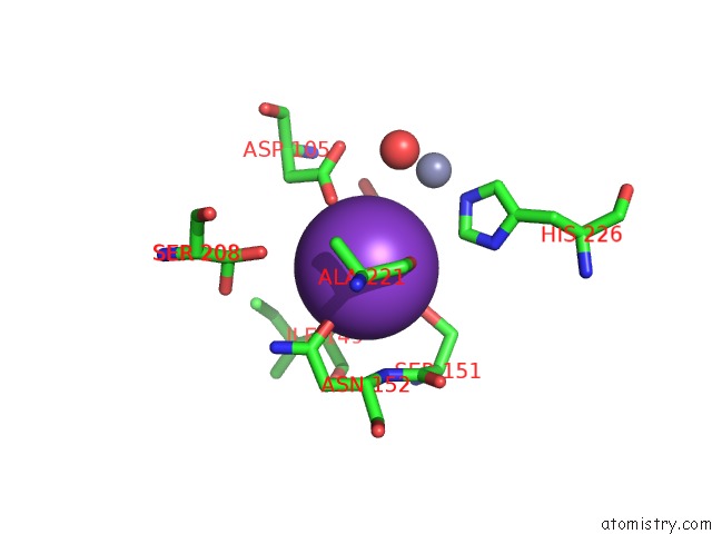
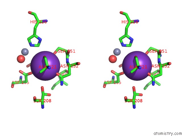
Potassium binding site 3 out of 4 in 4rfl
Go back to
Potassium binding site 3 out
of 4 in the Crystal Structure of G1PDH with Nadph From Methanocaldococcus Jannaschii
Mono view

Stereo pair view
Mono view
Stereo pair view
A full contact list of Potassium with other atoms in the K binding
site number 3 of Crystal Structure of G1PDH with Nadph From Methanocaldococcus Jannaschii within 5.0Å range:
|
Potassium binding site 4 out of 4 in 4rfl
Go back to
Potassium binding site 4 out
of 4 in the Crystal Structure of G1PDH with Nadph From Methanocaldococcus Jannaschii
Mono view
Stereo pair view
Mono view
Stereo pair view
A full contact list of Potassium with other atoms in the K binding
site number 4 of Crystal Structure of G1PDH with Nadph From Methanocaldococcus Jannaschii within 5.0Å range:
|
Reference:
V.Carbone,
L.R.Schofield,
Y.Zhang,
C.Sang,
D.Dey,
I.M.Hannus,
W.F.Martin,
A.J.Sutherland-Smith,
R.S.Ronimus.
Structure and Evolution of the Archaeal Lipid Synthesis Enzyme Sn-Glycerol-1-Phosphate Dehydrogenase. J.Biol.Chem. V. 290 21690 2015.
ISSN: ISSN 0021-9258
PubMed: 26175150
DOI: 10.1074/JBC.M115.647461
Page generated: Mon Aug 12 11:58:47 2024
ISSN: ISSN 0021-9258
PubMed: 26175150
DOI: 10.1074/JBC.M115.647461
Last articles
Zn in 9J0NZn in 9J0O
Zn in 9J0P
Zn in 9FJX
Zn in 9EKB
Zn in 9C0F
Zn in 9CAH
Zn in 9CH0
Zn in 9CH3
Zn in 9CH1