Potassium »
PDB 4qxg-4tog »
4r8i »
Potassium in PDB 4r8i: High Resolution Structure of A Mirror-Image Rna Oligonucleotide Aptamer in Complex with the Chemokine CCL2
Protein crystallography data
The structure of High Resolution Structure of A Mirror-Image Rna Oligonucleotide Aptamer in Complex with the Chemokine CCL2, PDB code: 4r8i
was solved by
D.Oberthuer,
J.Achenbach,
A.Gabdulkhakov,
S.Falke,
K.Buchner,
C.Maasch,
D.Rehders,
S.Klussmann,
C.Betzel,
with X-Ray Crystallography technique. A brief refinement statistics is given in the table below:
| Resolution Low / High (Å) | 77.01 / 2.05 |
| Space group | P 43 21 2 |
| Cell size a, b, c (Å), α, β, γ (°) | 108.912, 108.912, 34.812, 90.00, 90.00, 90.00 |
| R / Rfree (%) | 20 / 25 |
Other elements in 4r8i:
The structure of High Resolution Structure of A Mirror-Image Rna Oligonucleotide Aptamer in Complex with the Chemokine CCL2 also contains other interesting chemical elements:
| Strontium | (Sr) | 1 atom |
| Sodium | (Na) | 1 atom |
Potassium Binding Sites:
The binding sites of Potassium atom in the High Resolution Structure of A Mirror-Image Rna Oligonucleotide Aptamer in Complex with the Chemokine CCL2
(pdb code 4r8i). This binding sites where shown within
5.0 Angstroms radius around Potassium atom.
In total 5 binding sites of Potassium where determined in the High Resolution Structure of A Mirror-Image Rna Oligonucleotide Aptamer in Complex with the Chemokine CCL2, PDB code: 4r8i:
Jump to Potassium binding site number: 1; 2; 3; 4; 5;
In total 5 binding sites of Potassium where determined in the High Resolution Structure of A Mirror-Image Rna Oligonucleotide Aptamer in Complex with the Chemokine CCL2, PDB code: 4r8i:
Jump to Potassium binding site number: 1; 2; 3; 4; 5;
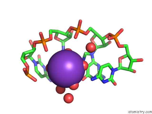
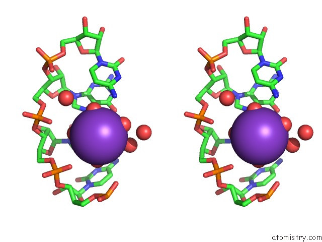
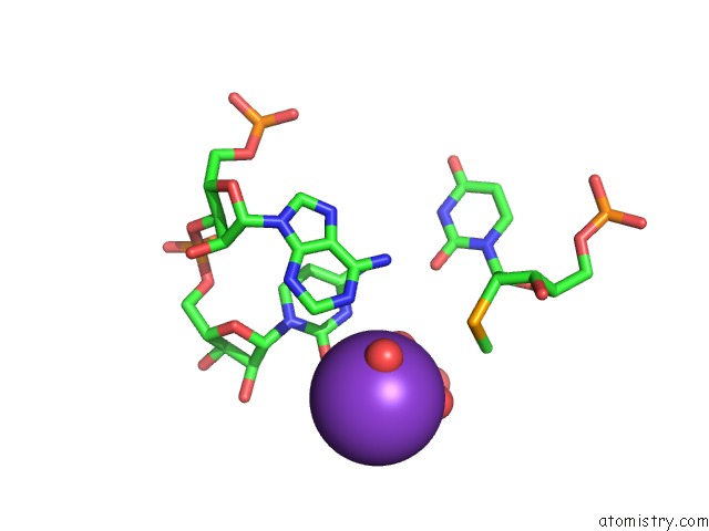
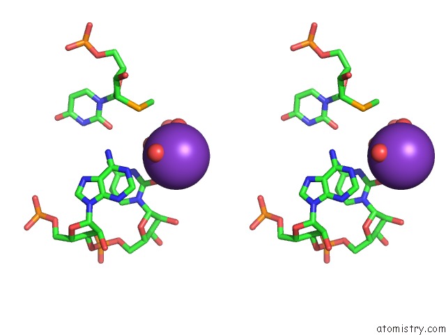
Potassium binding site 1 out of 5 in 4r8i
Go back to
Potassium binding site 1 out
of 5 in the High Resolution Structure of A Mirror-Image Rna Oligonucleotide Aptamer in Complex with the Chemokine CCL2
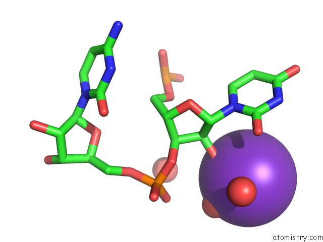
Mono view
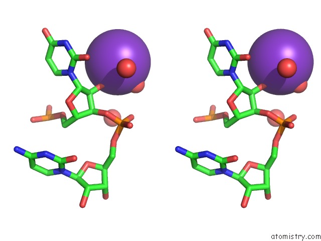
Stereo pair view
Mono view
Stereo pair view
A full contact list of Potassium with other atoms in the K binding
site number 1 of High Resolution Structure of A Mirror-Image Rna Oligonucleotide Aptamer in Complex with the Chemokine CCL2 within 5.0Å range:
|
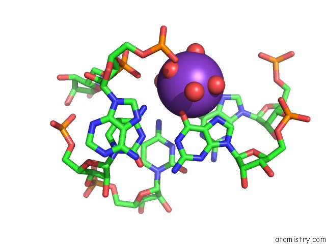
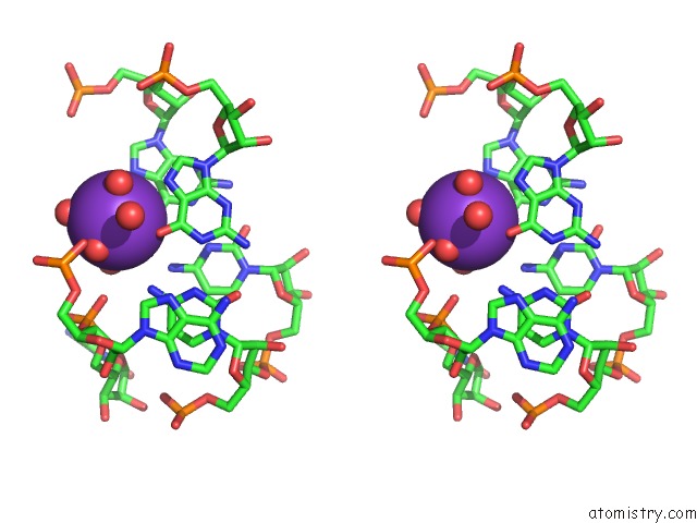
Potassium binding site 2 out of 5 in 4r8i
Go back to
Potassium binding site 2 out
of 5 in the High Resolution Structure of A Mirror-Image Rna Oligonucleotide Aptamer in Complex with the Chemokine CCL2
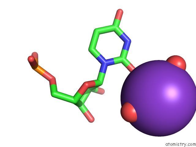
Mono view
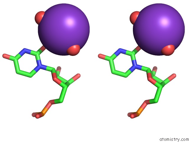
Stereo pair view
Mono view
Stereo pair view
A full contact list of Potassium with other atoms in the K binding
site number 2 of High Resolution Structure of A Mirror-Image Rna Oligonucleotide Aptamer in Complex with the Chemokine CCL2 within 5.0Å range:
|
Potassium binding site 3 out of 5 in 4r8i
Go back to
Potassium binding site 3 out
of 5 in the High Resolution Structure of A Mirror-Image Rna Oligonucleotide Aptamer in Complex with the Chemokine CCL2
Mono view
Stereo pair view
Mono view
Stereo pair view
A full contact list of Potassium with other atoms in the K binding
site number 3 of High Resolution Structure of A Mirror-Image Rna Oligonucleotide Aptamer in Complex with the Chemokine CCL2 within 5.0Å range:
|
Potassium binding site 4 out of 5 in 4r8i
Go back to
Potassium binding site 4 out
of 5 in the High Resolution Structure of A Mirror-Image Rna Oligonucleotide Aptamer in Complex with the Chemokine CCL2
Mono view
Stereo pair view
Mono view
Stereo pair view
A full contact list of Potassium with other atoms in the K binding
site number 4 of High Resolution Structure of A Mirror-Image Rna Oligonucleotide Aptamer in Complex with the Chemokine CCL2 within 5.0Å range:
|
Potassium binding site 5 out of 5 in 4r8i
Go back to
Potassium binding site 5 out
of 5 in the High Resolution Structure of A Mirror-Image Rna Oligonucleotide Aptamer in Complex with the Chemokine CCL2
Mono view
Stereo pair view
Mono view
Stereo pair view
A full contact list of Potassium with other atoms in the K binding
site number 5 of High Resolution Structure of A Mirror-Image Rna Oligonucleotide Aptamer in Complex with the Chemokine CCL2 within 5.0Å range:
|
Reference:
D.Oberthur,
J.Achenbach,
A.Gabdulkhakov,
K.Buchner,
C.Maasch,
S.Falke,
D.Rehders,
S.Klussmann,
C.Betzel.
Crystal Structure of A Mirror-Image L-Rna Aptamer (Spiegelmer) in Complex with the Natural L-Protein Target CCL2. Nat Commun V. 6 6923 2015.
ISSN: ESSN 2041-1723
PubMed: 25901662
DOI: 10.1038/NCOMMS7923
Page generated: Mon Aug 12 11:56:52 2024
ISSN: ESSN 2041-1723
PubMed: 25901662
DOI: 10.1038/NCOMMS7923
Last articles
Zn in 9J0NZn in 9J0O
Zn in 9J0P
Zn in 9FJX
Zn in 9EKB
Zn in 9C0F
Zn in 9CAH
Zn in 9CH0
Zn in 9CH3
Zn in 9CH1