Potassium in PDB 4r45: Racemic Crystal Structure of A Bimolecular Dna G-Quadruplex (P-1)
Protein crystallography data
The structure of Racemic Crystal Structure of A Bimolecular Dna G-Quadruplex (P-1), PDB code: 4r45
was solved by
P.K.Mandal,
G.W.Collie,
B.Kauffmann,
I.Huc,
with X-Ray Crystallography technique. A brief refinement statistics is given in the table below:
Resolution Low / High (Å)
|
25.02 /
1.90
|
Space group
|
P -1
|
Cell size a, b, c (Å), α, β, γ (°)
|
27.646,
28.300,
45.719,
104.50,
94.19,
113.42
|
R / Rfree (%)
|
28.6 /
34
|
Potassium Binding Sites:
The binding sites of Potassium atom in the Racemic Crystal Structure of A Bimolecular Dna G-Quadruplex (P-1)
(pdb code 4r45). This binding sites where shown within
5.0 Angstroms radius around Potassium atom.
In total 5 binding sites of Potassium where determined in the
Racemic Crystal Structure of A Bimolecular Dna G-Quadruplex (P-1), PDB code: 4r45:
Jump to Potassium binding site number:
1;
2;
3;
4;
5;
Potassium binding site 1 out
of 5 in 4r45
Go back to
Potassium Binding Sites List in 4r45
Potassium binding site 1 out
of 5 in the Racemic Crystal Structure of A Bimolecular Dna G-Quadruplex (P-1)
 Mono view
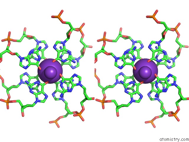
 Stereo pair view
|
A full contact list of Potassium with other atoms in the K binding
site number 1 of Racemic Crystal Structure of A Bimolecular Dna G-Quadruplex (P-1) within 5.0Å range:
probe
|
atom
|
residue
|
distance (Å)
|
B
|
Occ
|
A:K1101
b:6.5
occ:1.00
|
O6
|
A:DG1002
|
2.8
|
7.2
|
1.0
|
O6
|
A:DG1003
|
2.8
|
7.0
|
1.0
|
O6
|
A:DG1010
|
2.8
|
7.4
|
1.0
|
O6
|
B:DG2002
|
2.8
|
8.2
|
1.0
|
O6
|
B:DG2010
|
2.8
|
8.7
|
1.0
|
O6
|
B:DG2003
|
2.8
|
7.6
|
1.0
|
O6
|
B:DG2011
|
2.9
|
9.6
|
1.0
|
O6
|
A:DG1011
|
2.9
|
9.4
|
1.0
|
K
|
A:K1102
|
3.3
|
6.4
|
1.0
|
K
|
A:K1104
|
3.4
|
6.3
|
1.0
|
C6
|
A:DG1002
|
3.6
|
7.7
|
1.0
|
C6
|
B:DG2002
|
3.6
|
8.6
|
1.0
|
C6
|
A:DG1010
|
3.6
|
8.5
|
1.0
|
C6
|
B:DG2010
|
3.6
|
9.5
|
1.0
|
C6
|
A:DG1003
|
3.8
|
7.3
|
1.0
|
C6
|
B:DG2011
|
3.8
|
10.0
|
1.0
|
C6
|
B:DG2003
|
3.8
|
8.1
|
1.0
|
C6
|
A:DG1011
|
3.8
|
9.7
|
1.0
|
N1
|
A:DG1002
|
3.9
|
7.6
|
1.0
|
N1
|
B:DG2002
|
3.9
|
8.6
|
1.0
|
N1
|
B:DG2010
|
3.9
|
9.3
|
1.0
|
N1
|
A:DG1010
|
3.9
|
8.3
|
1.0
|
N1
|
B:DG2011
|
4.1
|
10.3
|
1.0
|
N1
|
A:DG1011
|
4.1
|
9.8
|
1.0
|
N1
|
A:DG1003
|
4.2
|
7.3
|
1.0
|
N1
|
B:DG2003
|
4.2
|
8.2
|
1.0
|
C5
|
A:DG1002
|
4.8
|
7.7
|
1.0
|
C5
|
B:DG2002
|
4.8
|
9.0
|
1.0
|
C5
|
A:DG1010
|
4.8
|
9.0
|
1.0
|
C5
|
B:DG2010
|
4.9
|
10.1
|
1.0
|
|
Potassium binding site 2 out
of 5 in 4r45
Go back to
Potassium Binding Sites List in 4r45
Potassium binding site 2 out
of 5 in the Racemic Crystal Structure of A Bimolecular Dna G-Quadruplex (P-1)
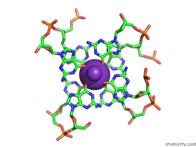
 Mono view
 Stereo pair view
|
A full contact list of Potassium with other atoms in the K binding
site number 2 of Racemic Crystal Structure of A Bimolecular Dna G-Quadruplex (P-1) within 5.0Å range:
probe
|
atom
|
residue
|
distance (Å)
|
B
|
Occ
|
A:K1102
b:6.4
occ:1.00
|
O6
|
B:DG2011
|
2.7
|
9.6
|
1.0
|
O6
|
B:DG2001
|
2.7
|
8.9
|
1.0
|
O6
|
A:DG1004
|
2.8
|
7.3
|
1.0
|
O6
|
A:DG1003
|
2.8
|
7.0
|
1.0
|
O6
|
B:DG2012
|
2.8
|
8.6
|
1.0
|
O6
|
A:DG1009
|
2.8
|
7.9
|
1.0
|
O6
|
A:DG1010
|
2.8
|
7.4
|
1.0
|
O6
|
B:DG2002
|
2.9
|
8.2
|
1.0
|
K
|
A:K1101
|
3.3
|
6.5
|
1.0
|
K
|
A:K1103
|
3.5
|
8.7
|
1.0
|
C6
|
B:DG2011
|
3.6
|
10.0
|
1.0
|
C6
|
A:DG1003
|
3.6
|
7.3
|
1.0
|
C6
|
B:DG2001
|
3.7
|
9.1
|
1.0
|
C6
|
A:DG1009
|
3.7
|
8.6
|
1.0
|
C6
|
A:DG1004
|
3.7
|
8.1
|
1.0
|
C6
|
A:DG1010
|
3.8
|
8.5
|
1.0
|
C6
|
B:DG2012
|
3.8
|
9.3
|
1.0
|
C6
|
B:DG2002
|
3.8
|
8.6
|
1.0
|
N1
|
A:DG1003
|
3.9
|
7.3
|
1.0
|
N1
|
B:DG2011
|
3.9
|
10.3
|
1.0
|
N1
|
B:DG2001
|
4.0
|
9.0
|
1.0
|
N1
|
A:DG1004
|
4.0
|
8.1
|
1.0
|
N1
|
A:DG1009
|
4.1
|
8.4
|
1.0
|
N1
|
B:DG2012
|
4.1
|
9.3
|
1.0
|
N1
|
A:DG1010
|
4.1
|
8.3
|
1.0
|
N1
|
B:DG2002
|
4.1
|
8.6
|
1.0
|
C5
|
B:DG2011
|
4.7
|
10.8
|
1.0
|
C5
|
A:DG1003
|
4.7
|
7.7
|
1.0
|
C5
|
B:DG2001
|
4.9
|
9.2
|
1.0
|
C5
|
A:DG1009
|
4.9
|
9.1
|
1.0
|
C5
|
A:DG1010
|
5.0
|
9.0
|
1.0
|
|
Potassium binding site 3 out
of 5 in 4r45
Go back to
Potassium Binding Sites List in 4r45
Potassium binding site 3 out
of 5 in the Racemic Crystal Structure of A Bimolecular Dna G-Quadruplex (P-1)
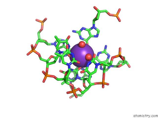
 Mono view
 Stereo pair view
|
A full contact list of Potassium with other atoms in the K binding
site number 3 of Racemic Crystal Structure of A Bimolecular Dna G-Quadruplex (P-1) within 5.0Å range:
probe
|
atom
|
residue
|
distance (Å)
|
B
|
Occ
|
A:K1103
b:8.7
occ:1.00
|
O2
|
A:DT1007
|
2.7
|
9.8
|
1.0
|
O
|
A:HOH1204
|
2.7
|
13.6
|
1.0
|
O6
|
B:DG2012
|
2.8
|
8.6
|
1.0
|
O2
|
A:DT1005
|
2.9
|
10.6
|
1.0
|
O6
|
A:DG1009
|
2.9
|
7.9
|
1.0
|
O6
|
A:DG1004
|
2.9
|
7.3
|
1.0
|
O
|
A:HOH1202
|
3.0
|
12.9
|
1.0
|
O6
|
B:DG2001
|
3.0
|
8.9
|
1.0
|
K
|
A:K1102
|
3.5
|
6.4
|
1.0
|
C6
|
B:DG2012
|
3.6
|
9.3
|
1.0
|
C2
|
A:DT1007
|
3.7
|
10.3
|
1.0
|
C6
|
A:DG1004
|
3.7
|
8.1
|
1.0
|
C6
|
A:DG1009
|
3.7
|
8.6
|
1.0
|
C2
|
A:DT1005
|
3.8
|
11.3
|
1.0
|
C6
|
B:DG2001
|
3.9
|
9.1
|
1.0
|
N3
|
A:DT1005
|
3.9
|
11.5
|
1.0
|
N1
|
B:DG2012
|
4.0
|
9.3
|
1.0
|
N3
|
A:DT1007
|
4.0
|
10.4
|
1.0
|
N1
|
A:DG1009
|
4.0
|
8.4
|
1.0
|
N1
|
B:DG2001
|
4.2
|
9.0
|
1.0
|
N1
|
A:DG1004
|
4.2
|
8.1
|
1.0
|
O2
|
A:DT1006
|
4.3
|
11.8
|
1.0
|
C5
|
A:DG1004
|
4.7
|
8.5
|
1.0
|
C5
|
B:DG2012
|
4.8
|
10.3
|
1.0
|
N1
|
A:DT1007
|
4.9
|
11.0
|
1.0
|
C5
|
A:DG1009
|
5.0
|
9.1
|
1.0
|
C2
|
A:DT1006
|
5.0
|
12.6
|
1.0
|
O4'
|
A:DT1008
|
5.0
|
11.3
|
1.0
|
|
Potassium binding site 4 out
of 5 in 4r45
Go back to
Potassium Binding Sites List in 4r45
Potassium binding site 4 out
of 5 in the Racemic Crystal Structure of A Bimolecular Dna G-Quadruplex (P-1)
 Mono view
 Stereo pair view
|
A full contact list of Potassium with other atoms in the K binding
site number 4 of Racemic Crystal Structure of A Bimolecular Dna G-Quadruplex (P-1) within 5.0Å range:
probe
|
atom
|
residue
|
distance (Å)
|
B
|
Occ
|
A:K1104
b:6.3
occ:1.00
|
O6
|
A:DG1001
|
2.7
|
8.0
|
1.0
|
O6
|
A:DG1011
|
2.8
|
9.4
|
1.0
|
O6
|
B:DG2003
|
2.8
|
7.6
|
1.0
|
O6
|
B:DG2004
|
2.8
|
9.0
|
1.0
|
O6
|
B:DG2009
|
2.8
|
8.6
|
1.0
|
O6
|
A:DG1012
|
2.9
|
9.0
|
1.0
|
O6
|
B:DG2010
|
2.9
|
8.7
|
1.0
|
O6
|
A:DG1002
|
2.9
|
7.2
|
1.0
|
K
|
A:K1101
|
3.4
|
6.5
|
1.0
|
K
|
B:K2101
|
3.5
|
9.1
|
1.0
|
C6
|
B:DG2003
|
3.6
|
8.1
|
1.0
|
C6
|
A:DG1011
|
3.6
|
9.7
|
1.0
|
C6
|
A:DG1001
|
3.6
|
8.2
|
1.0
|
C6
|
B:DG2004
|
3.7
|
9.4
|
1.0
|
C6
|
B:DG2009
|
3.7
|
9.4
|
1.0
|
C6
|
B:DG2010
|
3.8
|
9.5
|
1.0
|
C6
|
A:DG1012
|
3.8
|
10.0
|
1.0
|
C6
|
A:DG1002
|
3.8
|
7.7
|
1.0
|
N1
|
B:DG2003
|
3.9
|
8.2
|
1.0
|
N1
|
A:DG1011
|
3.9
|
9.8
|
1.0
|
N1
|
A:DG1001
|
4.0
|
8.1
|
1.0
|
N1
|
B:DG2004
|
4.0
|
9.5
|
1.0
|
N1
|
B:DG2009
|
4.1
|
9.4
|
1.0
|
N1
|
A:DG1012
|
4.1
|
10.2
|
1.0
|
N1
|
B:DG2010
|
4.1
|
9.3
|
1.0
|
N1
|
A:DG1002
|
4.1
|
7.6
|
1.0
|
C5
|
B:DG2003
|
4.7
|
8.4
|
1.0
|
C5
|
A:DG1011
|
4.7
|
10.6
|
1.0
|
C5
|
A:DG1001
|
4.9
|
8.4
|
1.0
|
C5
|
B:DG2009
|
4.9
|
9.9
|
1.0
|
|
Potassium binding site 5 out
of 5 in 4r45
Go back to
Potassium Binding Sites List in 4r45
Potassium binding site 5 out
of 5 in the Racemic Crystal Structure of A Bimolecular Dna G-Quadruplex (P-1)
 Mono view
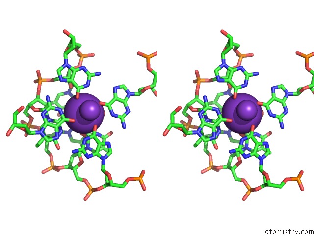
 Stereo pair view
|
A full contact list of Potassium with other atoms in the K binding
site number 5 of Racemic Crystal Structure of A Bimolecular Dna G-Quadruplex (P-1) within 5.0Å range:
probe
|
atom
|
residue
|
distance (Å)
|
B
|
Occ
|
B:K2101
b:9.1
occ:1.00
|
O2
|
B:DT2007
|
2.7
|
11.4
|
1.0
|
O
|
B:HOH2212
|
2.8
|
12.8
|
1.0
|
O6
|
A:DG1012
|
2.8
|
9.0
|
1.0
|
O6
|
B:DG2004
|
2.9
|
9.0
|
1.0
|
O6
|
B:DG2009
|
2.9
|
8.6
|
1.0
|
O2
|
B:DT2005
|
2.9
|
11.2
|
1.0
|
O
|
B:HOH2204
|
2.9
|
14.9
|
1.0
|
O6
|
A:DG1001
|
3.0
|
8.0
|
1.0
|
K
|
A:K1104
|
3.5
|
6.3
|
1.0
|
C6
|
A:DG1012
|
3.6
|
10.0
|
1.0
|
C2
|
B:DT2007
|
3.6
|
11.7
|
1.0
|
C6
|
B:DG2004
|
3.7
|
9.4
|
1.0
|
C6
|
B:DG2009
|
3.8
|
9.4
|
1.0
|
C2
|
B:DT2005
|
3.8
|
12.3
|
1.0
|
C6
|
A:DG1001
|
3.9
|
8.2
|
1.0
|
N3
|
B:DT2005
|
3.9
|
12.6
|
1.0
|
N3
|
B:DT2007
|
3.9
|
11.7
|
1.0
|
N1
|
A:DG1012
|
4.0
|
10.2
|
1.0
|
N1
|
B:DG2009
|
4.0
|
9.4
|
1.0
|
N1
|
A:DG1001
|
4.2
|
8.1
|
1.0
|
N1
|
B:DG2004
|
4.2
|
9.5
|
1.0
|
O2
|
B:DT2006
|
4.3
|
13.7
|
1.0
|
C5
|
B:DG2004
|
4.7
|
9.8
|
1.0
|
C5
|
A:DG1012
|
4.8
|
11.2
|
1.0
|
N1
|
B:DT2007
|
4.9
|
12.1
|
1.0
|
C2
|
B:DT2006
|
5.0
|
14.8
|
1.0
|
O4'
|
B:DT2008
|
5.0
|
14.0
|
1.0
|
|
Reference:
P.K.Mandal,
G.W.Collie,
B.Kauffmann,
I.Huc.
Racemic Dna Crystallography. Angew.Chem.Int.Ed.Engl. 2014.
ISSN: ESSN 1521-3773
PubMed: 25358289
DOI: 10.1002/ANIE.201409014
Page generated: Mon Aug 12 11:55:31 2024
|