Potassium »
PDB 4qxg-4tog »
4r33 »
Potassium in PDB 4r33: X-Ray Structure of the Tryptophan Lyase Nosl with Tryptophan and S- Adenosyl-L-Homocysteine Bound
Protein crystallography data
The structure of X-Ray Structure of the Tryptophan Lyase Nosl with Tryptophan and S- Adenosyl-L-Homocysteine Bound, PDB code: 4r33
was solved by
Y.Nicolet,
L.Zeppieri,
P.Amara,
J.-C.Fontecilla-Camps,
with X-Ray Crystallography technique. A brief refinement statistics is given in the table below:
| Resolution Low / High (Å) | 47.23 / 1.78 |
| Space group | P 1 2 1 |
| Cell size a, b, c (Å), α, β, γ (°) | 94.720, 47.230, 114.360, 90.00, 108.71, 90.00 |
| R / Rfree (%) | 15.9 / 18.1 |
Other elements in 4r33:
The structure of X-Ray Structure of the Tryptophan Lyase Nosl with Tryptophan and S- Adenosyl-L-Homocysteine Bound also contains other interesting chemical elements:
| Iron | (Fe) | 8 atoms |
| Chlorine | (Cl) | 2 atoms |
| Sodium | (Na) | 1 atom |
Potassium Binding Sites:
The binding sites of Potassium atom in the X-Ray Structure of the Tryptophan Lyase Nosl with Tryptophan and S- Adenosyl-L-Homocysteine Bound
(pdb code 4r33). This binding sites where shown within
5.0 Angstroms radius around Potassium atom.
In total 2 binding sites of Potassium where determined in the X-Ray Structure of the Tryptophan Lyase Nosl with Tryptophan and S- Adenosyl-L-Homocysteine Bound, PDB code: 4r33:
Jump to Potassium binding site number: 1; 2;
In total 2 binding sites of Potassium where determined in the X-Ray Structure of the Tryptophan Lyase Nosl with Tryptophan and S- Adenosyl-L-Homocysteine Bound, PDB code: 4r33:
Jump to Potassium binding site number: 1; 2;
Potassium binding site 1 out of 2 in 4r33
Go back to
Potassium binding site 1 out
of 2 in the X-Ray Structure of the Tryptophan Lyase Nosl with Tryptophan and S- Adenosyl-L-Homocysteine Bound
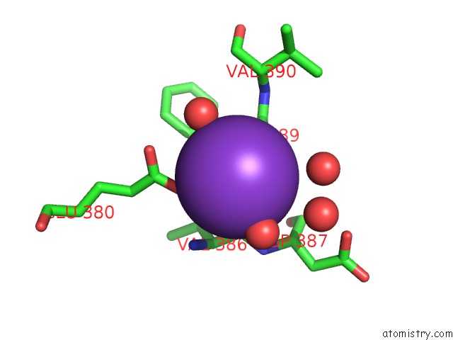
Mono view
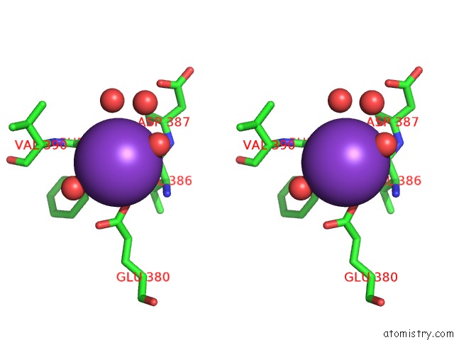
Stereo pair view
Mono view
Stereo pair view
A full contact list of Potassium with other atoms in the K binding
site number 1 of X-Ray Structure of the Tryptophan Lyase Nosl with Tryptophan and S- Adenosyl-L-Homocysteine Bound within 5.0Å range:
|
Potassium binding site 2 out of 2 in 4r33
Go back to
Potassium binding site 2 out
of 2 in the X-Ray Structure of the Tryptophan Lyase Nosl with Tryptophan and S- Adenosyl-L-Homocysteine Bound
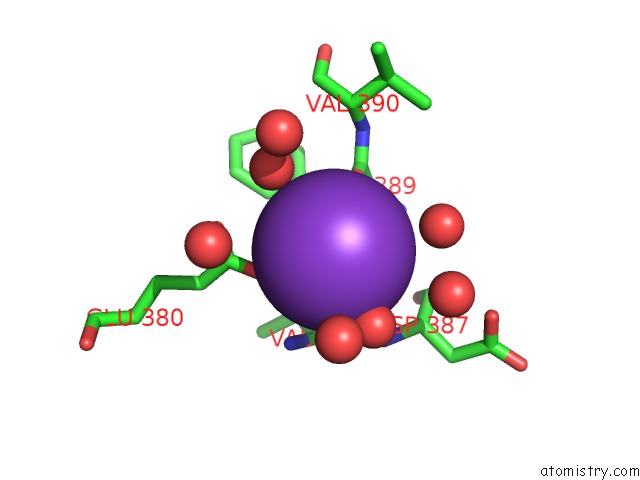
Mono view
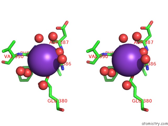
Stereo pair view
Mono view
Stereo pair view
A full contact list of Potassium with other atoms in the K binding
site number 2 of X-Ray Structure of the Tryptophan Lyase Nosl with Tryptophan and S- Adenosyl-L-Homocysteine Bound within 5.0Å range:
|
Reference:
Y.Nicolet,
L.Zeppieri,
P.Amara,
J.C.Fontecilla-Camps.
Crystal Structure of Tryptophan Lyase (Nosl): Evidence For Radical Formation at the Amino Group of Tryptophan. Angew.Chem.Int.Ed.Engl. V. 53 11840 2014.
ISSN: ISSN 1433-7851
PubMed: 25196319
DOI: 10.1002/ANIE.201407320
Page generated: Mon Aug 12 11:55:31 2024
ISSN: ISSN 1433-7851
PubMed: 25196319
DOI: 10.1002/ANIE.201407320
Last articles
Zn in 9J0NZn in 9J0O
Zn in 9J0P
Zn in 9FJX
Zn in 9EKB
Zn in 9C0F
Zn in 9CAH
Zn in 9CH0
Zn in 9CH3
Zn in 9CH1