Potassium »
PDB 4mlz-4pjo »
4o6h »
Potassium in PDB 4o6h: 2.8A Crystal Structure of Lymphocytic Choriomeningitis Virus Nucleoprotein C-Terminal Domain
Protein crystallography data
The structure of 2.8A Crystal Structure of Lymphocytic Choriomeningitis Virus Nucleoprotein C-Terminal Domain, PDB code: 4o6h
was solved by
B.R.West,
K.M.Hastie,
E.O.Saphire,
with X-Ray Crystallography technique. A brief refinement statistics is given in the table below:
| Resolution Low / High (Å) | 47.26 / 2.80 |
| Space group | P 1 21 1 |
| Cell size a, b, c (Å), α, β, γ (°) | 92.856, 94.403, 145.124, 90.00, 102.30, 90.00 |
| R / Rfree (%) | 22.7 / 26.1 |
Other elements in 4o6h:
The structure of 2.8A Crystal Structure of Lymphocytic Choriomeningitis Virus Nucleoprotein C-Terminal Domain also contains other interesting chemical elements:
| Magnesium | (Mg) | 4 atoms |
| Zinc | (Zn) | 8 atoms |
Potassium Binding Sites:
The binding sites of Potassium atom in the 2.8A Crystal Structure of Lymphocytic Choriomeningitis Virus Nucleoprotein C-Terminal Domain
(pdb code 4o6h). This binding sites where shown within
5.0 Angstroms radius around Potassium atom.
In total 4 binding sites of Potassium where determined in the 2.8A Crystal Structure of Lymphocytic Choriomeningitis Virus Nucleoprotein C-Terminal Domain, PDB code: 4o6h:
Jump to Potassium binding site number: 1; 2; 3; 4;
In total 4 binding sites of Potassium where determined in the 2.8A Crystal Structure of Lymphocytic Choriomeningitis Virus Nucleoprotein C-Terminal Domain, PDB code: 4o6h:
Jump to Potassium binding site number: 1; 2; 3; 4;
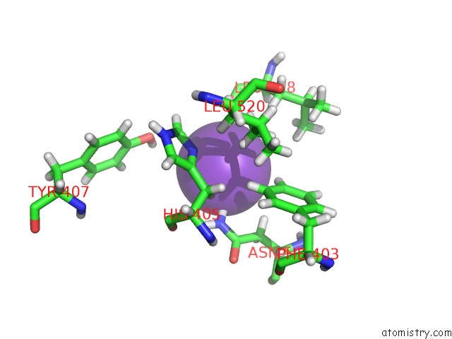
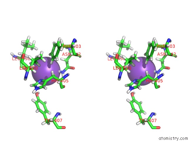
Potassium binding site 1 out of 4 in 4o6h
Go back to
Potassium binding site 1 out
of 4 in the 2.8A Crystal Structure of Lymphocytic Choriomeningitis Virus Nucleoprotein C-Terminal Domain
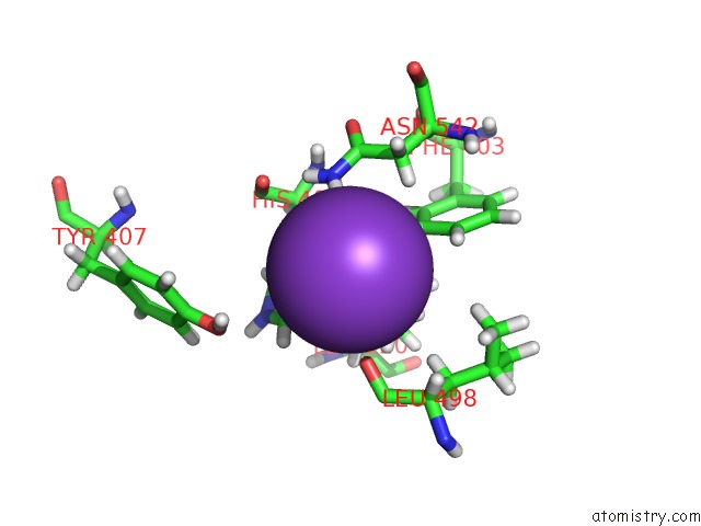
Mono view
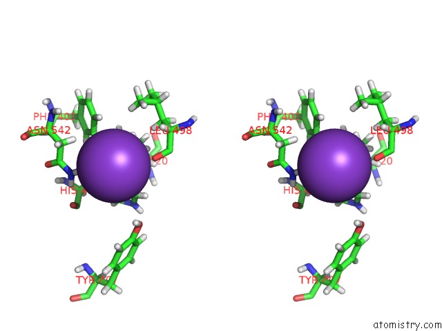
Stereo pair view
Mono view
Stereo pair view
A full contact list of Potassium with other atoms in the K binding
site number 1 of 2.8A Crystal Structure of Lymphocytic Choriomeningitis Virus Nucleoprotein C-Terminal Domain within 5.0Å range:
|
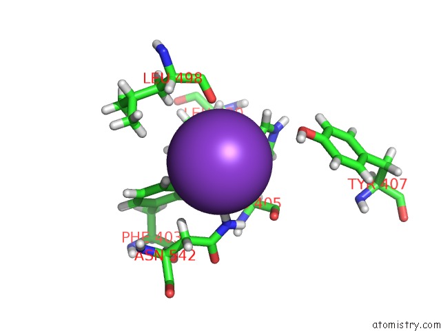
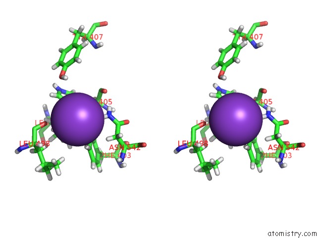
Potassium binding site 2 out of 4 in 4o6h
Go back to
Potassium binding site 2 out
of 4 in the 2.8A Crystal Structure of Lymphocytic Choriomeningitis Virus Nucleoprotein C-Terminal Domain
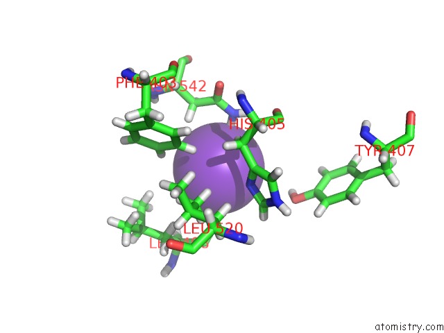
Mono view
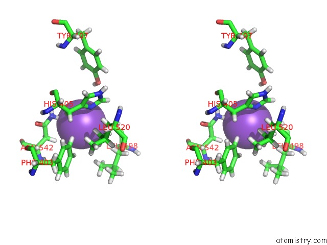
Stereo pair view
Mono view
Stereo pair view
A full contact list of Potassium with other atoms in the K binding
site number 2 of 2.8A Crystal Structure of Lymphocytic Choriomeningitis Virus Nucleoprotein C-Terminal Domain within 5.0Å range:
|
Potassium binding site 3 out of 4 in 4o6h
Go back to
Potassium binding site 3 out
of 4 in the 2.8A Crystal Structure of Lymphocytic Choriomeningitis Virus Nucleoprotein C-Terminal Domain
Mono view
Stereo pair view
Mono view
Stereo pair view
A full contact list of Potassium with other atoms in the K binding
site number 3 of 2.8A Crystal Structure of Lymphocytic Choriomeningitis Virus Nucleoprotein C-Terminal Domain within 5.0Å range:
|
Potassium binding site 4 out of 4 in 4o6h
Go back to
Potassium binding site 4 out
of 4 in the 2.8A Crystal Structure of Lymphocytic Choriomeningitis Virus Nucleoprotein C-Terminal Domain
Mono view
Stereo pair view
Mono view
Stereo pair view
A full contact list of Potassium with other atoms in the K binding
site number 4 of 2.8A Crystal Structure of Lymphocytic Choriomeningitis Virus Nucleoprotein C-Terminal Domain within 5.0Å range:
|
Reference:
B.R.West,
K.M.Hastie,
E.O.Saphire.
Structure of the Lcmv Nucleoprotein Provides A Template For Understanding Arenavirus Replication and Immunosuppression. Acta Crystallogr.,Sect.D V. 70 1764 2014.
ISSN: ISSN 0907-4449
PubMed: 24914986
DOI: 10.1107/S1399004714007883
Page generated: Sat Aug 9 07:33:14 2025
ISSN: ISSN 0907-4449
PubMed: 24914986
DOI: 10.1107/S1399004714007883
Last articles
Mg in 4AC0Mg in 4AAS
Mg in 4AAR
Mg in 4AAQ
Mg in 4AAB
Mg in 4A95
Mg in 4A8W
Mg in 4A8Y
Mg in 4A93
Mg in 4A8S