Atomistry » Potassium » PDB 4l4d-4mkk » 4lzb
Atomistry »
  Potassium »
    PDB 4l4d-4mkk »
      4lzb »

Potassium in PDB 4lzb: Uracil Binding Pocket in Vaccinia Virus Uracil Dna Glycosylase

Enzymatic activity of Uracil Binding Pocket in Vaccinia Virus Uracil Dna Glycosylase

All present enzymatic activity of Uracil Binding Pocket in Vaccinia Virus Uracil Dna Glycosylase:
3.2.2.27;

Protein crystallography data

The structure of Uracil Binding Pocket in Vaccinia Virus Uracil Dna Glycosylase, PDB code: 4lzb was solved by N.Schormann, D.Chattopadhyay, with X-Ray Crystallography technique. A brief refinement statistics is given in the table below:

Resolution Low / High (Å) 49.64 / 2.03
Space group P 21 21 21
Cell size a, b, c (Å), α, β, γ (°) 93.470, 114.045, 302.516, 90.00, 90.00, 90.00
R / Rfree (%) 21.3 / 24.6

Other elements in 4lzb:

The structure of Uracil Binding Pocket in Vaccinia Virus Uracil Dna Glycosylase also contains other interesting chemical elements:

Chlorine (Cl) 14 atoms

Potassium Binding Sites:

The binding sites of Potassium atom in the Uracil Binding Pocket in Vaccinia Virus Uracil Dna Glycosylase (pdb code 4lzb). This binding sites where shown within 5.0 Angstroms radius around Potassium atom.
In total 10 binding sites of Potassium where determined in the Uracil Binding Pocket in Vaccinia Virus Uracil Dna Glycosylase, PDB code: 4lzb:
Jump to Potassium binding site number: 1; 2; 3; 4; 5; 6; 7; 8; 9; 10;

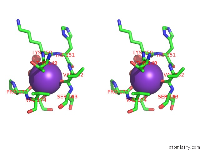
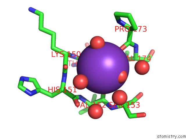
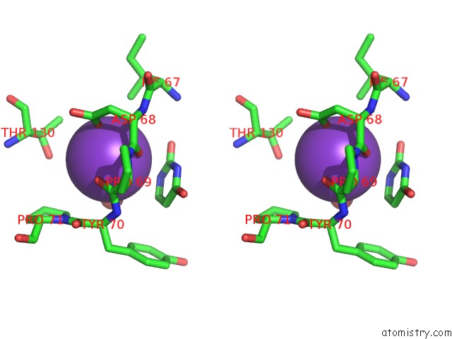
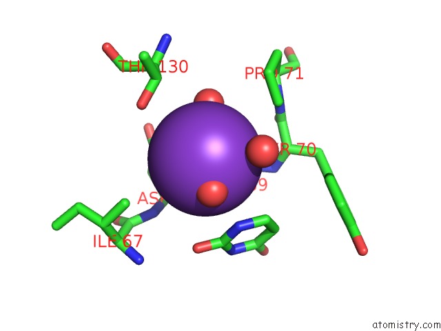
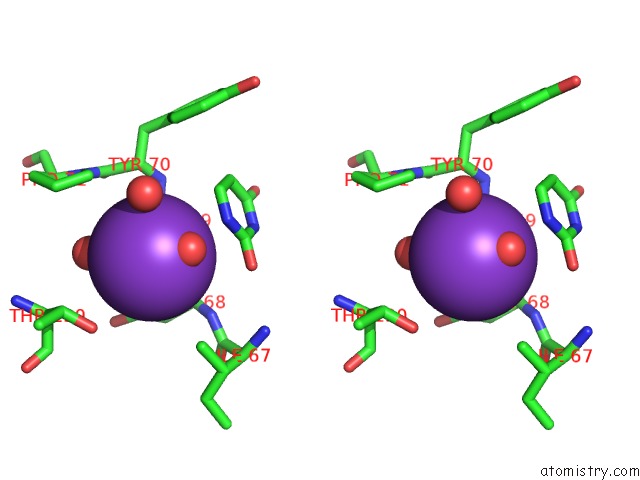
Potassium binding site 1 out of 10 in 4lzb

Go back to Potassium Binding Sites List in 4lzb
Potassium binding site 1 out of 10 in the Uracil Binding Pocket in Vaccinia Virus Uracil Dna Glycosylase


Mono view


Stereo pair view

A full contact list of Potassium with other atoms in the K binding site number 1 of Uracil Binding Pocket in Vaccinia Virus Uracil Dna Glycosylase within 5.0Å range:
probe atom residue distance (Å) B Occ
A:K302

b:42.0
occ:1.00
O A:HOH495 2.7 40.1 1.0
OD1 A:ASP68 2.7 20.7 1.0
O A:HOH546 2.7 31.3 1.0
O A:HOH631 2.8 41.4 1.0
O A:ASP68 3.0 16.3 1.0
O A:PRO69 3.0 17.8 1.0
N1 A:URA308 3.5 25.4 1.0
C A:ASP68 3.7 16.9 1.0
C A:PRO69 3.7 17.9 1.0
CG A:ASP68 3.8 19.8 1.0
C6 A:URA308 3.9 24.7 1.0
C2 A:URA308 4.0 25.3 1.0
CG2 A:ILE67 4.2 17.6 1.0
CG2 A:THR130 4.3 31.4 1.0
O2 A:URA308 4.3 25.5 1.0
N A:TYR70 4.3 18.3 1.0
OG1 A:THR130 4.3 32.5 1.0
N A:PRO69 4.4 17.3 1.0
N A:ASP68 4.4 16.1 1.0
CD A:PRO71 4.4 22.6 1.0
CA A:TYR70 4.5 18.4 1.0
CA A:ASP68 4.5 17.2 1.0
CA A:PRO69 4.6 17.4 1.0
C5 A:URA308 4.6 24.3 1.0
OD2 A:ASP68 4.6 21.6 1.0
N3 A:URA308 4.6 24.9 1.0
CB A:ASP68 4.8 18.1 1.0
CB A:THR130 4.9 31.2 1.0
C4 A:URA308 4.9 23.9 1.0
N A:PRO71 5.0 21.8 1.0
C A:TYR70 5.0 20.0 1.0

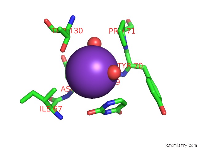
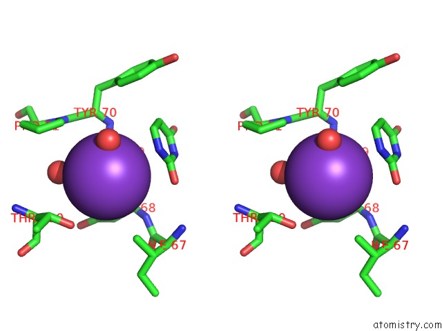
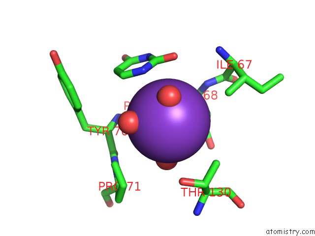
Potassium binding site 2 out of 10 in 4lzb

Go back to Potassium Binding Sites List in 4lzb
Potassium binding site 2 out of 10 in the Uracil Binding Pocket in Vaccinia Virus Uracil Dna Glycosylase


Mono view


Stereo pair view

A full contact list of Potassium with other atoms in the K binding site number 2 of Uracil Binding Pocket in Vaccinia Virus Uracil Dna Glycosylase within 5.0Å range:
probe atom residue distance (Å) B Occ
B:K304

b:16.8
occ:1.00
O B:VAL152 2.6 12.8 1.0
O B:HOH420 2.7 21.9 1.0
O B:THR149 2.7 16.0 1.0
O B:PRO173 2.8 18.6 1.0
O2 B:EDO309 3.0 25.5 1.0
C B:THR149 3.8 16.5 1.0
C B:VAL152 3.8 14.3 1.0
O B:LYS150 3.9 15.6 1.0
C B:LYS150 4.0 16.7 1.0
C B:PRO173 4.0 18.7 1.0
C2 B:EDO309 4.1 23.8 1.0
O B:HOH465 4.2 19.2 1.0
CB B:VAL174 4.3 17.1 1.0
CA B:LYS150 4.3 17.3 1.0
CA B:VAL174 4.4 17.6 1.0
N B:HIS151 4.5 16.6 1.0
N B:VAL152 4.5 15.3 1.0
CG2 B:VAL174 4.5 17.4 1.0
N B:LYS150 4.5 17.0 1.0
C B:HIS151 4.6 16.5 1.0
CA B:SER153 4.6 14.1 0.5
CA B:SER153 4.6 14.5 0.5
N B:VAL174 4.7 17.9 1.0
N B:SER153 4.7 14.2 0.5
N B:SER153 4.7 13.9 0.5
CA B:THR149 4.8 16.2 1.0
CA B:VAL152 4.8 14.6 1.0
O B:HIS151 4.8 16.5 1.0
CA B:HIS151 5.0 17.0 1.0

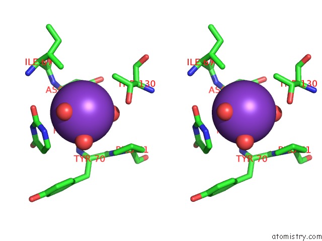
Potassium binding site 3 out of 10 in 4lzb

Go back to Potassium Binding Sites List in 4lzb
Potassium binding site 3 out of 10 in the Uracil Binding Pocket in Vaccinia Virus Uracil Dna Glycosylase


Mono view


Stereo pair view

A full contact list of Potassium with other atoms in the K binding site number 3 of Uracil Binding Pocket in Vaccinia Virus Uracil Dna Glycosylase within 5.0Å range:
probe atom residue distance (Å) B Occ
B:K305

b:38.9
occ:1.00
OD1 B:ASP68 2.7 18.4 1.0
O B:HOH596 2.8 43.8 1.0
O B:PRO69 2.9 18.6 1.0
O B:ASP68 3.0 16.0 1.0
O B:HOH467 3.3 34.2 1.0
C B:ASP68 3.6 16.1 1.0
C B:PRO69 3.7 18.3 1.0
N1 B:URA312 3.7 21.0 1.0
OG1 B:THR130 3.8 29.5 1.0
CG B:ASP68 3.8 17.4 1.0
C2 B:URA312 4.0 20.7 1.0
C6 B:URA312 4.1 21.2 1.0
O2 B:URA312 4.2 20.5 1.0
CG2 B:ILE67 4.3 15.8 1.0
N B:PRO69 4.3 17.0 1.0
N B:ASP68 4.4 14.5 1.0
CD B:PRO71 4.4 24.5 1.0
N B:TYR70 4.4 20.0 1.0
CB B:THR130 4.4 28.3 1.0
CA B:ASP68 4.4 15.6 1.0
CA B:PRO69 4.5 17.6 1.0
CA B:TYR70 4.5 20.4 1.0
OD2 B:ASP68 4.6 18.3 1.0
N3 B:URA312 4.7 20.0 1.0
CB B:ASP68 4.7 16.3 1.0
C5 B:URA312 4.8 20.6 1.0
N B:PRO71 4.8 23.8 1.0
C B:TYR70 4.9 21.7 1.0

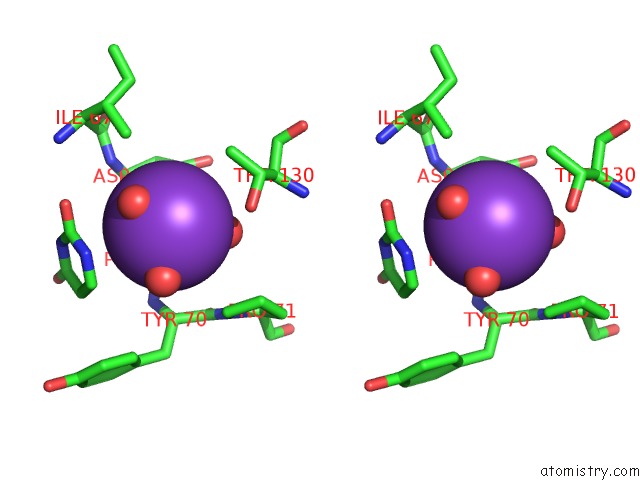
Potassium binding site 4 out of 10 in 4lzb

Go back to Potassium Binding Sites List in 4lzb
Potassium binding site 4 out of 10 in the Uracil Binding Pocket in Vaccinia Virus Uracil Dna Glycosylase


Mono view


Stereo pair view

A full contact list of Potassium with other atoms in the K binding site number 4 of Uracil Binding Pocket in Vaccinia Virus Uracil Dna Glycosylase within 5.0Å range:
probe atom residue distance (Å) B Occ
C:K301

b:42.7
occ:1.00
O C:HOH492 2.6 36.3 1.0
O C:HOH493 2.6 47.7 1.0
OD1 C:ASP68 2.7 32.2 1.0
O C:PRO69 3.0 28.1 1.0
O C:ASP68 3.0 26.7 1.0
OG1 C:THR130 3.7 41.6 1.0
N1 C:URA303 3.7 30.1 1.0
C C:ASP68 3.8 27.3 1.0
CG C:ASP68 3.8 30.8 1.0
C C:PRO69 3.8 28.2 1.0
C6 C:URA303 4.0 30.2 1.0
C2 C:URA303 4.1 30.0 1.0
CG2 C:THR130 4.1 39.5 1.0
CG2 C:ILE67 4.3 27.9 1.0
N C:ASP68 4.4 27.6 1.0
O2 C:URA303 4.4 29.7 1.0
OD2 C:ASP68 4.4 32.2 1.0
N C:TYR70 4.5 29.1 1.0
CB C:THR130 4.5 39.3 1.0
CA C:ASP68 4.5 27.6 1.0
N C:PRO69 4.5 26.8 1.0
CD C:PRO71 4.5 33.2 1.0
CA C:TYR70 4.5 29.4 1.0
CA C:PRO69 4.7 27.8 1.0
C5 C:URA303 4.7 29.3 1.0
CB C:ASP68 4.7 29.1 1.0
N3 C:URA303 4.7 29.0 1.0
N C:PRO71 4.9 32.7 1.0
C C:TYR70 5.0 30.6 1.0
C4 C:URA303 5.0 28.6 1.0

Potassium binding site 5 out of 10 in 4lzb

Go back to Potassium Binding Sites List in 4lzb
Potassium binding site 5 out of 10 in the Uracil Binding Pocket in Vaccinia Virus Uracil Dna Glycosylase


Mono view


Stereo pair view

A full contact list of Potassium with other atoms in the K binding site number 5 of Uracil Binding Pocket in Vaccinia Virus Uracil Dna Glycosylase within 5.0Å range:
probe atom residue distance (Å) B Occ
E:K302

b:19.1
occ:1.00
O E:HOH568 2.3 27.9 1.0
O E:THR149 2.6 17.5 1.0
O E:VAL152 2.6 16.3 1.0
O E:PRO173 2.7 18.5 1.0
O E:HOH567 2.9 34.1 1.0
O A:HOH529 3.8 38.7 1.0
C E:THR149 3.8 17.6 1.0
C E:VAL152 3.8 16.4 1.0
C E:PRO173 3.9 18.9 1.0
O E:LYS150 3.9 20.1 1.0
C E:LYS150 4.0 19.6 1.0
O E:HOH511 4.1 28.0 1.0
O E:HOH570 4.2 36.3 1.0
CA E:LYS150 4.3 20.0 1.0
CB E:VAL174 4.3 17.7 1.0
N E:HIS151 4.3 18.9 1.0
N E:VAL152 4.4 17.3 1.0
C E:HIS151 4.4 18.1 1.0
CA E:VAL174 4.4 17.7 1.0
CG2 E:VAL174 4.5 17.8 1.0
N E:LYS150 4.5 18.7 1.0
N E:SER153 4.6 16.0 1.0
N E:VAL174 4.6 18.2 1.0
CA E:SER153 4.6 15.1 1.0
O E:HIS151 4.7 17.6 1.0
CA E:VAL152 4.7 16.8 1.0
CA E:THR149 4.8 16.7 1.0
O E:HOH514 4.9 35.2 1.0
CA E:HIS151 4.9 18.9 1.0

Potassium binding site 6 out of 10 in 4lzb

Go back to Potassium Binding Sites List in 4lzb
Potassium binding site 6 out of 10 in the Uracil Binding Pocket in Vaccinia Virus Uracil Dna Glycosylase


Mono view


Stereo pair view

A full contact list of Potassium with other atoms in the K binding site number 6 of Uracil Binding Pocket in Vaccinia Virus Uracil Dna Glycosylase within 5.0Å range:
probe atom residue distance (Å) B Occ
D:K301

b:51.2
occ:1.00
O D:HOH495 2.7 38.9 1.0
OD1 D:ASP68 2.8 37.7 1.0
O D:ASP68 3.0 31.4 1.0
O D:PRO69 3.1 30.6 1.0
N1 D:URA305 3.6 29.9 1.0
C D:ASP68 3.8 31.4 1.0
C D:PRO69 3.8 30.3 1.0
C2 D:URA305 3.9 29.6 1.0
OG1 D:THR130 3.9 46.6 1.0
CG D:ASP68 3.9 36.2 1.0
C6 D:URA305 4.0 30.1 1.0
O2 D:URA305 4.1 29.7 1.0
CG2 D:THR130 4.2 43.1 1.0
CG2 D:ILE67 4.3 29.6 1.0
N D:ASP68 4.4 29.4 1.0
N D:TYR70 4.4 30.3 1.0
CD D:PRO71 4.5 32.6 1.0
CA D:ASP68 4.5 31.6 1.0
N3 D:URA305 4.5 28.8 1.0
CA D:TYR70 4.5 29.0 1.0
N D:PRO69 4.6 30.3 1.0
C5 D:URA305 4.7 29.3 1.0
CA D:PRO69 4.7 30.2 1.0
CB D:THR130 4.7 43.4 1.0
OD2 D:ASP68 4.7 37.8 1.0
CB D:ASP68 4.9 33.7 1.0
C4 D:URA305 4.9 28.5 1.0

Potassium binding site 7 out of 10 in 4lzb

Go back to Potassium Binding Sites List in 4lzb
Potassium binding site 7 out of 10 in the Uracil Binding Pocket in Vaccinia Virus Uracil Dna Glycosylase


Mono view


Stereo pair view

A full contact list of Potassium with other atoms in the K binding site number 7 of Uracil Binding Pocket in Vaccinia Virus Uracil Dna Glycosylase within 5.0Å range:
probe atom residue distance (Å) B Occ
E:K303

b:40.7
occ:1.00
OD1 E:ASP68 2.6 23.9 1.0
O E:HOH571 2.6 51.3 1.0
O E:HOH572 2.8 40.6 1.0
O E:HOH573 2.8 39.2 1.0
O E:PRO69 3.0 21.1 1.0
O E:ASP68 3.1 19.6 1.0
N1 E:URA307 3.7 21.8 1.0
C E:ASP68 3.7 20.6 1.0
CG E:ASP68 3.8 23.8 1.0
C E:PRO69 3.8 21.3 1.0
C2 E:URA307 4.0 21.5 1.0
OG1 E:THR130 4.1 34.1 1.0
C6 E:URA307 4.1 21.6 1.0
CG2 E:THR130 4.2 32.5 1.0
O2 E:URA307 4.2 22.4 1.0
CG2 E:ILE67 4.3 18.7 1.0
N E:ASP68 4.4 19.9 1.0
CA E:ASP68 4.4 20.9 1.0
N E:PRO69 4.4 20.5 1.0
N E:TYR70 4.5 23.3 1.0
CD E:PRO71 4.5 29.9 1.0
OD2 E:ASP68 4.5 27.1 1.0
CA E:TYR70 4.5 25.1 1.0
N3 E:URA307 4.6 21.1 1.0
CA E:PRO69 4.6 21.1 1.0
CB E:ASP68 4.7 21.9 1.0
CB E:THR130 4.7 32.3 1.0
C5 E:URA307 4.8 21.3 1.0
N E:PRO71 4.9 29.3 1.0
C E:TYR70 4.9 27.2 1.0
C4 E:URA307 5.0 20.2 1.0

Potassium binding site 8 out of 10 in 4lzb

Go back to Potassium Binding Sites List in 4lzb
Potassium binding site 8 out of 10 in the Uracil Binding Pocket in Vaccinia Virus Uracil Dna Glycosylase


Mono view


Stereo pair view

A full contact list of Potassium with other atoms in the K binding site number 8 of Uracil Binding Pocket in Vaccinia Virus Uracil Dna Glycosylase within 5.0Å range:
probe atom residue distance (Å) B Occ
F:K302

b:41.5
occ:1.00
OD1 F:ASP68 2.8 22.3 1.0
O F:HOH584 2.9 35.8 1.0
O F:ASP68 3.0 21.4 1.0
O F:PRO69 3.0 21.0 1.0
O F:HOH466 3.4 31.6 1.0
OG1 F:THR130 3.6 34.1 1.0
N1 F:URA305 3.7 23.0 1.0
C F:ASP68 3.8 21.6 1.0
C F:PRO69 3.8 21.9 1.0
C2 F:URA305 3.9 22.6 1.0
CG F:ASP68 4.0 22.7 1.0
O2 F:URA305 4.0 23.2 1.0
O F:HOH520 4.1 41.7 1.0
C6 F:URA305 4.1 23.1 1.0
O F:HOH401 4.3 33.9 1.0
N F:ASP68 4.4 20.2 1.0
CG2 F:ILE67 4.4 20.6 1.0
N3 F:URA305 4.5 22.0 1.0
N F:TYR70 4.5 22.5 1.0
CA F:ASP68 4.5 21.2 1.0
CB F:THR130 4.5 32.9 1.0
N F:PRO69 4.5 21.8 1.0
CD F:PRO71 4.6 26.2 1.0
CA F:TYR70 4.6 22.9 1.0
CA F:PRO69 4.7 22.1 1.0
C5 F:URA305 4.7 22.2 1.0
OD2 F:ASP68 4.7 23.0 1.0
C4 F:URA305 4.8 21.3 1.0
CB F:ASP68 4.9 22.0 1.0

Potassium binding site 9 out of 10 in 4lzb

Go back to Potassium Binding Sites List in 4lzb
Potassium binding site 9 out of 10 in the Uracil Binding Pocket in Vaccinia Virus Uracil Dna Glycosylase


Mono view


Stereo pair view

A full contact list of Potassium with other atoms in the K binding site number 9 of Uracil Binding Pocket in Vaccinia Virus Uracil Dna Glycosylase within 5.0Å range:
probe atom residue distance (Å) B Occ
J:K303

b:43.7
occ:1.00
O J:HOH506 2.5 51.1 1.0
O J:HOH507 2.7 42.1 1.0
O J:HOH505 2.7 38.9 1.0
OD1 J:ASP68 2.8 30.0 1.0
O J:PRO69 3.1 25.4 1.0
O J:ASP68 3.3 25.7 1.0
OG1 J:THR130 3.6 39.5 1.0
N1 J:URA301 3.7 31.5 1.0
C J:PRO69 3.9 25.8 1.0
CG J:ASP68 4.0 29.4 1.0
C J:ASP68 4.0 25.6 1.0
C6 J:URA301 4.0 31.7 1.0
CG2 J:THR130 4.1 37.8 1.0
C2 J:URA301 4.1 30.9 1.0
CG2 J:ILE67 4.3 24.4 1.0
O2 J:URA301 4.4 29.8 1.0
CD J:PRO71 4.4 30.6 1.0
CB J:THR130 4.4 38.0 1.0
N J:TYR70 4.5 27.1 1.0
CA J:TYR70 4.6 27.3 1.0
N J:ASP68 4.6 26.1 1.0
OD2 J:ASP68 4.7 29.6 1.0
N J:PRO69 4.7 24.5 1.0
CA J:ASP68 4.7 26.1 1.0
C5 J:URA301 4.8 31.8 1.0
N3 J:URA301 4.8 31.7 1.0
CA J:PRO69 4.8 24.7 1.0
N J:PRO71 4.9 29.5 1.0
CB J:ASP68 5.0 27.4 1.0
C J:TYR70 5.0 28.6 1.0
CG J:PRO71 5.0 30.8 1.0

Potassium binding site 10 out of 10 in 4lzb

Go back to Potassium Binding Sites List in 4lzb
Potassium binding site 10 out of 10 in the Uracil Binding Pocket in Vaccinia Virus Uracil Dna Glycosylase


Mono view


Stereo pair view

A full contact list of Potassium with other atoms in the K binding site number 10 of Uracil Binding Pocket in Vaccinia Virus Uracil Dna Glycosylase within 5.0Å range:
probe atom residue distance (Å) B Occ
G:K303

b:34.1
occ:1.00
O G:HOH540 2.4 27.5 1.0
OD1 G:ASP68 2.7 25.5 1.0
O G:HOH539 3.0 34.4 1.0
O G:PRO69 3.0 23.1 1.0
O G:ASP68 3.1 22.4 1.0
O G:HOH510 3.3 43.0 1.0
N1 G:URA306 3.6 18.0 1.0
C G:ASP68 3.8 22.9 1.0
CG G:ASP68 3.9 25.6 1.0
C G:PRO69 3.9 23.2 1.0
OG1 G:THR130 4.0 32.9 1.0
C2 G:URA306 4.1 18.3 1.0
C6 G:URA306 4.1 18.5 1.0
O2 G:URA306 4.3 18.6 1.0
CG2 G:ILE67 4.4 24.0 1.0
CD G:PRO71 4.4 28.5 1.0
N G:ASP68 4.4 22.4 1.0
CB G:THR130 4.5 31.4 1.0
N G:TYR70 4.5 23.8 1.0
CA G:ASP68 4.5 23.1 1.0
N G:PRO69 4.6 23.3 1.0
CA G:TYR70 4.6 24.2 1.0
OD2 G:ASP68 4.6 27.9 1.0
CA G:PRO69 4.8 23.4 1.0
N3 G:URA306 4.8 18.6 1.0
CB G:ASP68 4.8 24.1 1.0
C5 G:URA306 4.8 18.3 1.0
N G:PRO71 4.9 27.9 1.0
C G:TYR70 5.0 25.8 1.0

Reference:

N.Schormann, S.Banerjee, R.Ricciardi, D.Chattopadhyay. Structure of the Uracil Complex of Vaccinia Virus Uracil Dna Glycosylase. Acta Crystallogr.,Sect.F V. 69 1328 2013.
ISSN: ESSN 1744-3091
PubMed: 24316823
DOI: 10.1107/S1744309113030613
Page generated: Sun Dec 13 23:44:45 2020

Last articles

Zn in 8WB0
Zn in 8WAX
Zn in 8WAU
Zn in 8WAZ
Zn in 8WAY
Zn in 8WAV
Zn in 8WAW
Zn in 8WAT
Zn in 8W7M
Zn in 8WD3
© Copyright 2008-2020 by atomistry.com
Home   |    Site Map   |    Copyright   |    Contact us   |    Privacy