Potassium »
PDB 4l4d-4mkk »
4l4g »
Potassium in PDB 4l4g: Structure of Cyanide and Camphor Bound P450CAM Mutant L358P/K178G
Enzymatic activity of Structure of Cyanide and Camphor Bound P450CAM Mutant L358P/K178G
All present enzymatic activity of Structure of Cyanide and Camphor Bound P450CAM Mutant L358P/K178G:
1.14.15.1;
1.14.15.1;
Protein crystallography data
The structure of Structure of Cyanide and Camphor Bound P450CAM Mutant L358P/K178G, PDB code: 4l4g
was solved by
D.Batabyal,
H.Li,
T.L.Poulos,
with X-Ray Crystallography technique. A brief refinement statistics is given in the table below:
| Resolution Low / High (Å) | 35.88 / 1.55 |
| Space group | P 1 21 1 |
| Cell size a, b, c (Å), α, β, γ (°) | 57.072, 58.790, 57.072, 90.00, 105.02, 90.00 |
| R / Rfree (%) | 17.3 / 21.2 |
Other elements in 4l4g:
The structure of Structure of Cyanide and Camphor Bound P450CAM Mutant L358P/K178G also contains other interesting chemical elements:
| Iron | (Fe) | 1 atom |
Potassium Binding Sites:
The binding sites of Potassium atom in the Structure of Cyanide and Camphor Bound P450CAM Mutant L358P/K178G
(pdb code 4l4g). This binding sites where shown within
5.0 Angstroms radius around Potassium atom.
In total 3 binding sites of Potassium where determined in the Structure of Cyanide and Camphor Bound P450CAM Mutant L358P/K178G, PDB code: 4l4g:
Jump to Potassium binding site number: 1; 2; 3;
In total 3 binding sites of Potassium where determined in the Structure of Cyanide and Camphor Bound P450CAM Mutant L358P/K178G, PDB code: 4l4g:
Jump to Potassium binding site number: 1; 2; 3;
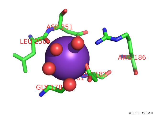
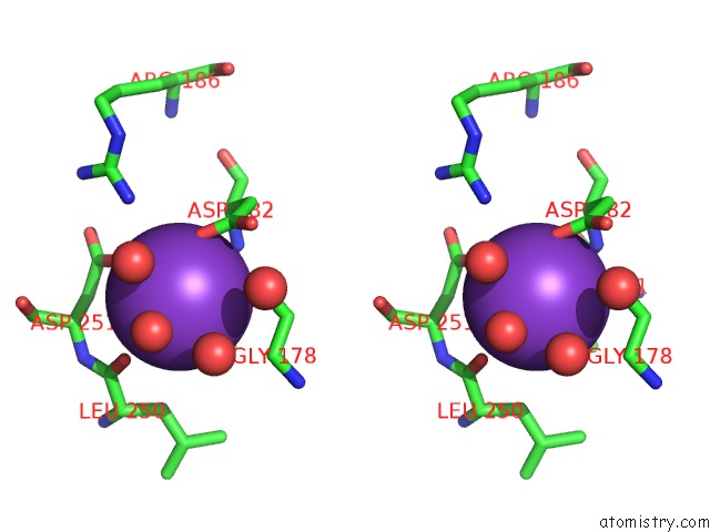
Potassium binding site 1 out of 3 in 4l4g
Go back to
Potassium binding site 1 out
of 3 in the Structure of Cyanide and Camphor Bound P450CAM Mutant L358P/K178G
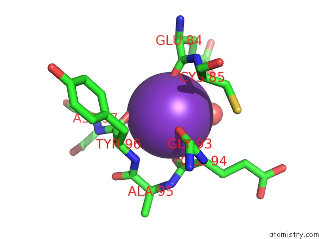
Mono view
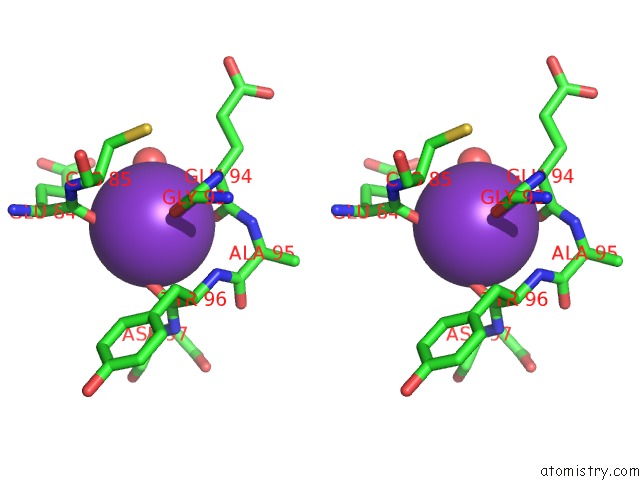
Stereo pair view
Mono view
Stereo pair view
A full contact list of Potassium with other atoms in the K binding
site number 1 of Structure of Cyanide and Camphor Bound P450CAM Mutant L358P/K178G within 5.0Å range:
|
Potassium binding site 2 out of 3 in 4l4g
Go back to
Potassium binding site 2 out
of 3 in the Structure of Cyanide and Camphor Bound P450CAM Mutant L358P/K178G
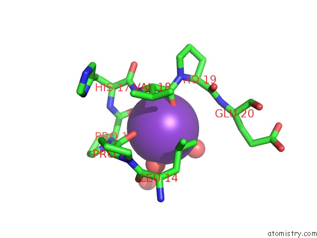
Mono view
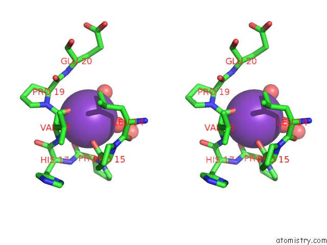
Stereo pair view
Mono view
Stereo pair view
A full contact list of Potassium with other atoms in the K binding
site number 2 of Structure of Cyanide and Camphor Bound P450CAM Mutant L358P/K178G within 5.0Å range:
|
Potassium binding site 3 out of 3 in 4l4g
Go back to
Potassium binding site 3 out
of 3 in the Structure of Cyanide and Camphor Bound P450CAM Mutant L358P/K178G
Mono view
Stereo pair view
Mono view
Stereo pair view
A full contact list of Potassium with other atoms in the K binding
site number 3 of Structure of Cyanide and Camphor Bound P450CAM Mutant L358P/K178G within 5.0Å range:
|
Reference:
D.Batabyal,
H.Li,
T.L.Poulos.
Synergistic Effects of Mutations in Cytochrome P450CAM Designed to Mimic CYP101D1. Biochemistry V. 52 5396 2013.
ISSN: ISSN 0006-2960
PubMed: 23865948
DOI: 10.1021/BI400676D
Page generated: Mon Aug 12 11:20:00 2024
ISSN: ISSN 0006-2960
PubMed: 23865948
DOI: 10.1021/BI400676D
Last articles
Zn in 9J0NZn in 9J0O
Zn in 9J0P
Zn in 9FJX
Zn in 9EKB
Zn in 9C0F
Zn in 9CAH
Zn in 9CH0
Zn in 9CH3
Zn in 9CH1