Potassium »
PDB 4ikr-4k8t »
4k8p »
Potassium in PDB 4k8p: Crystal Structure of Probable Sugar Kinase Protein From Rhizobium Etli Cfn 42 Complexed with 2-Ethylbenzyl Alcohol
Protein crystallography data
The structure of Crystal Structure of Probable Sugar Kinase Protein From Rhizobium Etli Cfn 42 Complexed with 2-Ethylbenzyl Alcohol, PDB code: 4k8p
was solved by
V.N.Malashkevich,
R.Bhosle,
R.Toro,
B.Hillerich,
A.Gizzi,
S.Garforth,
A.Kar,
M.K.Chan,
J.Lafluer,
H.Patel,
B.Matikainen,
S.Chamala,
S.Lim,
A.Celikgil,
G.Villegas,
B.Evans,
J.Love,
A.Fiser,
K.Khafizov,
R.Seidel,
J.B.Bonanno,
S.C.Almo,
New York Structural Genomics Researchconsortium (Nysgrc),
with X-Ray Crystallography technique. A brief refinement statistics is given in the table below:
| Resolution Low / High (Å) | 40.09 / 1.50 |
| Space group | P 1 21 1 |
| Cell size a, b, c (Å), α, β, γ (°) | 51.429, 80.818, 82.591, 90.00, 97.85, 90.00 |
| R / Rfree (%) | 18.5 / 23.6 |
Potassium Binding Sites:
The binding sites of Potassium atom in the Crystal Structure of Probable Sugar Kinase Protein From Rhizobium Etli Cfn 42 Complexed with 2-Ethylbenzyl Alcohol
(pdb code 4k8p). This binding sites where shown within
5.0 Angstroms radius around Potassium atom.
In total 2 binding sites of Potassium where determined in the Crystal Structure of Probable Sugar Kinase Protein From Rhizobium Etli Cfn 42 Complexed with 2-Ethylbenzyl Alcohol, PDB code: 4k8p:
Jump to Potassium binding site number: 1; 2;
In total 2 binding sites of Potassium where determined in the Crystal Structure of Probable Sugar Kinase Protein From Rhizobium Etli Cfn 42 Complexed with 2-Ethylbenzyl Alcohol, PDB code: 4k8p:
Jump to Potassium binding site number: 1; 2;
Potassium binding site 1 out of 2 in 4k8p
Go back to
Potassium binding site 1 out
of 2 in the Crystal Structure of Probable Sugar Kinase Protein From Rhizobium Etli Cfn 42 Complexed with 2-Ethylbenzyl Alcohol
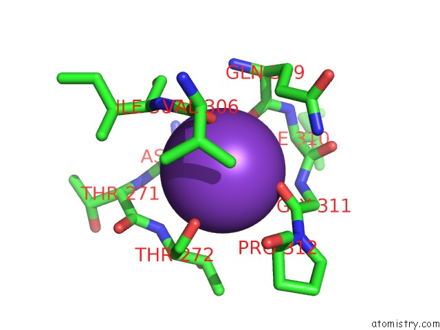
Mono view
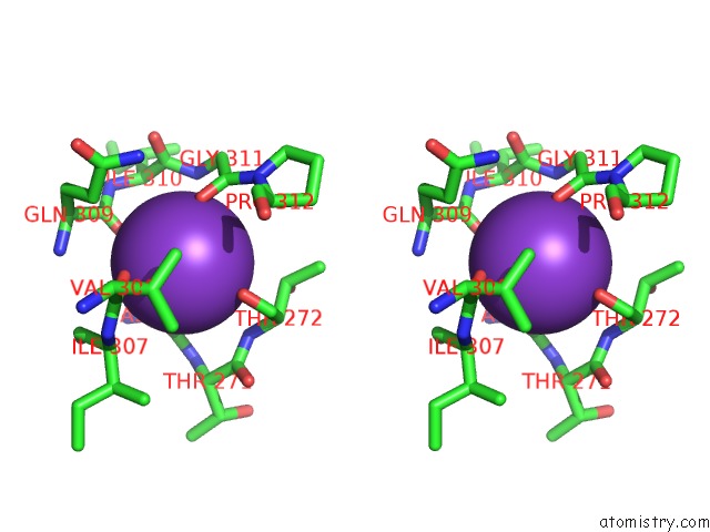
Stereo pair view
Mono view
Stereo pair view
A full contact list of Potassium with other atoms in the K binding
site number 1 of Crystal Structure of Probable Sugar Kinase Protein From Rhizobium Etli Cfn 42 Complexed with 2-Ethylbenzyl Alcohol within 5.0Å range:
|
Potassium binding site 2 out of 2 in 4k8p
Go back to
Potassium binding site 2 out
of 2 in the Crystal Structure of Probable Sugar Kinase Protein From Rhizobium Etli Cfn 42 Complexed with 2-Ethylbenzyl Alcohol
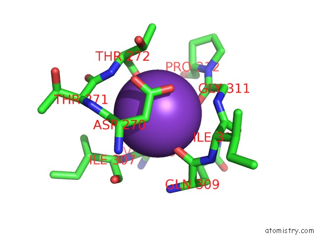
Mono view
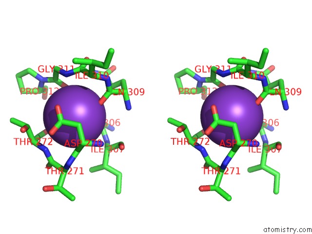
Stereo pair view
Mono view
Stereo pair view
A full contact list of Potassium with other atoms in the K binding
site number 2 of Crystal Structure of Probable Sugar Kinase Protein From Rhizobium Etli Cfn 42 Complexed with 2-Ethylbenzyl Alcohol within 5.0Å range:
|
Reference:
V.N.Malashkevich,
R.Bhosle,
R.Toro,
B.Hillerich,
A.Gizzi,
S.Garforth,
A.Kar,
M.K.Chan,
J.Lafluer,
H.Patel,
B.Matikainen,
S.Chamala,
S.Lim,
A.Celikgil,
G.Villegas,
B.Evans,
J.Love,
A.Fiser,
K.Khafizov,
R.Seidel,
J.B.Bonanno,
S.C.Almo.
Crystal Structure of Probable Sugar Kinase Protein From Rhizobium Etli Cfn 42 Complexed with 2-Ethylbenzyl Alcohol To Be Published.
Page generated: Mon Aug 12 11:08:09 2024
Last articles
Zn in 9JPJZn in 9JP7
Zn in 9JPK
Zn in 9JPL
Zn in 9GN6
Zn in 9GN7
Zn in 9GKU
Zn in 9GKW
Zn in 9GKX
Zn in 9GL0