Potassium »
PDB 4gx1-4ije »
4hyo »
Potassium in PDB 4hyo: Crystal Structure of Mthk Pore
Protein crystallography data
The structure of Crystal Structure of Mthk Pore, PDB code: 4hyo
was solved by
D.J.Posson,
J.G.Mccoy,
C.M.Nimigean,
with X-Ray Crystallography technique. A brief refinement statistics is given in the table below:
| Resolution Low / High (Å) | 44.86 / 1.65 |
| Space group | P 1 |
| Cell size a, b, c (Å), α, β, γ (°) | 44.035, 63.452, 63.477, 90.03, 89.99, 89.99 |
| R / Rfree (%) | 16.4 / 18.1 |
Potassium Binding Sites:
The binding sites of Potassium atom in the Crystal Structure of Mthk Pore
(pdb code 4hyo). This binding sites where shown within
5.0 Angstroms radius around Potassium atom.
In total 8 binding sites of Potassium where determined in the Crystal Structure of Mthk Pore, PDB code: 4hyo:
Jump to Potassium binding site number: 1; 2; 3; 4; 5; 6; 7; 8;
In total 8 binding sites of Potassium where determined in the Crystal Structure of Mthk Pore, PDB code: 4hyo:
Jump to Potassium binding site number: 1; 2; 3; 4; 5; 6; 7; 8;
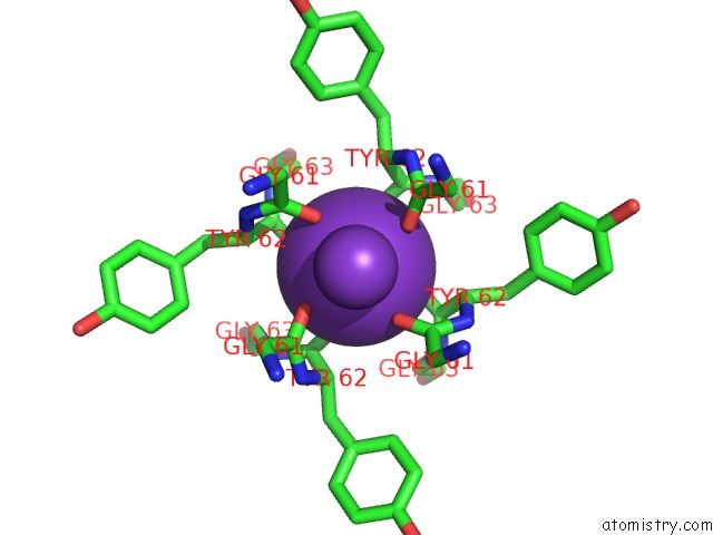
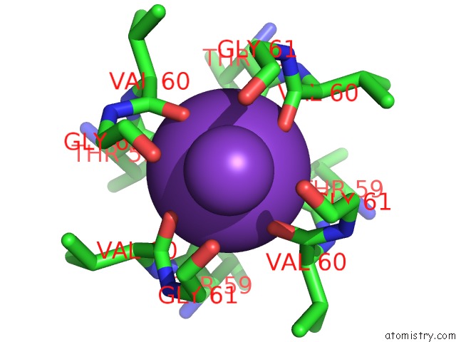
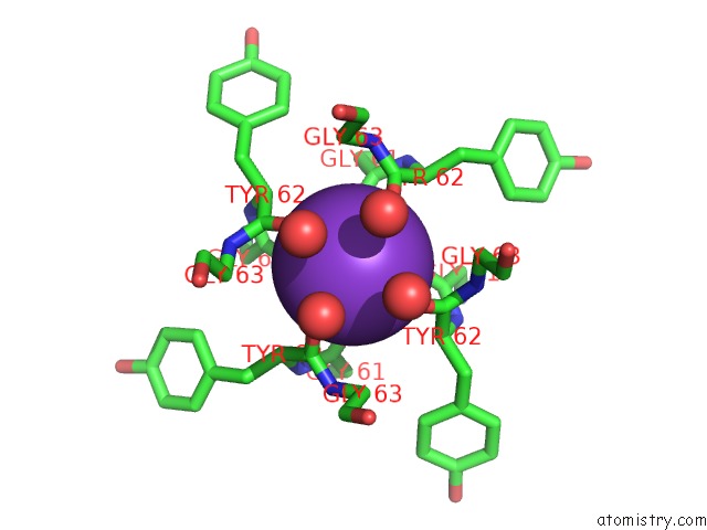
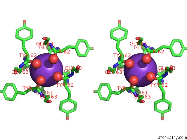
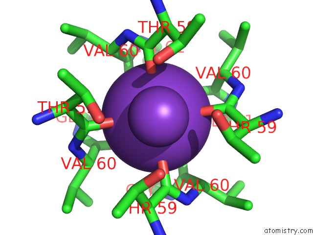
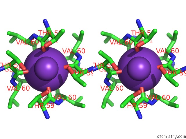
Potassium binding site 1 out of 8 in 4hyo
Go back to
Potassium binding site 1 out
of 8 in the Crystal Structure of Mthk Pore
Mono view
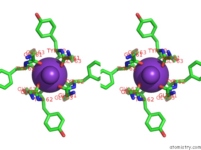
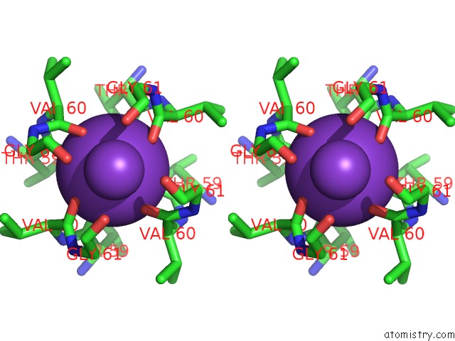
Stereo pair view
Mono view
Stereo pair view
A full contact list of Potassium with other atoms in the K binding
site number 1 of Crystal Structure of Mthk Pore within 5.0Å range:
|
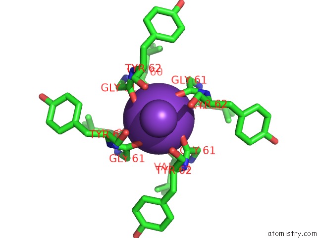
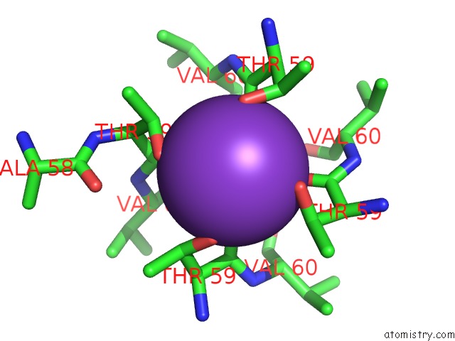
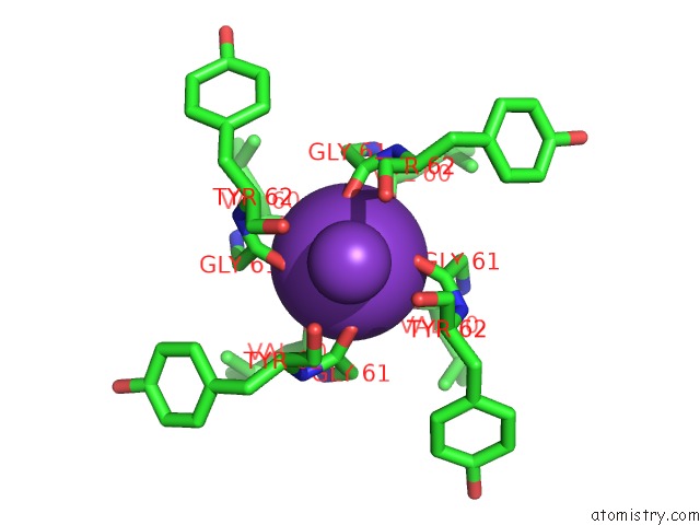
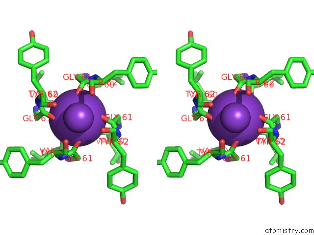
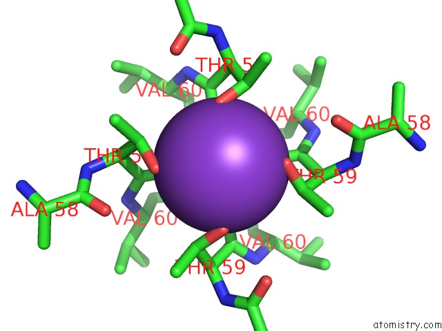
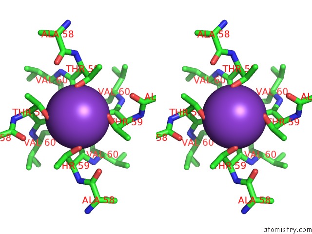
Potassium binding site 2 out of 8 in 4hyo
Go back to
Potassium binding site 2 out
of 8 in the Crystal Structure of Mthk Pore
Mono view
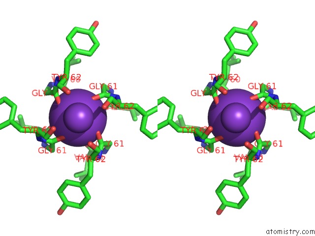
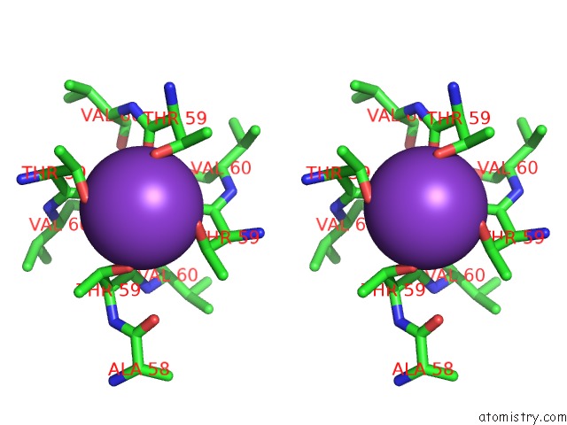
Stereo pair view
Mono view
Stereo pair view
A full contact list of Potassium with other atoms in the K binding
site number 2 of Crystal Structure of Mthk Pore within 5.0Å range:
|
Potassium binding site 3 out of 8 in 4hyo
Go back to
Potassium binding site 3 out
of 8 in the Crystal Structure of Mthk Pore
Mono view
Stereo pair view
Mono view
Stereo pair view
A full contact list of Potassium with other atoms in the K binding
site number 3 of Crystal Structure of Mthk Pore within 5.0Å range:
|
Potassium binding site 4 out of 8 in 4hyo
Go back to
Potassium binding site 4 out
of 8 in the Crystal Structure of Mthk Pore
Mono view
Stereo pair view
Mono view
Stereo pair view
A full contact list of Potassium with other atoms in the K binding
site number 4 of Crystal Structure of Mthk Pore within 5.0Å range:
|
Potassium binding site 5 out of 8 in 4hyo
Go back to
Potassium binding site 5 out
of 8 in the Crystal Structure of Mthk Pore
Mono view
Stereo pair view
Mono view
Stereo pair view
A full contact list of Potassium with other atoms in the K binding
site number 5 of Crystal Structure of Mthk Pore within 5.0Å range:
|
Potassium binding site 6 out of 8 in 4hyo
Go back to
Potassium binding site 6 out
of 8 in the Crystal Structure of Mthk Pore
Mono view
Stereo pair view
Mono view
Stereo pair view
A full contact list of Potassium with other atoms in the K binding
site number 6 of Crystal Structure of Mthk Pore within 5.0Å range:
|
Potassium binding site 7 out of 8 in 4hyo
Go back to
Potassium binding site 7 out
of 8 in the Crystal Structure of Mthk Pore
Mono view
Stereo pair view
Mono view
Stereo pair view
A full contact list of Potassium with other atoms in the K binding
site number 7 of Crystal Structure of Mthk Pore within 5.0Å range:
|
Potassium binding site 8 out of 8 in 4hyo
Go back to
Potassium binding site 8 out
of 8 in the Crystal Structure of Mthk Pore
Mono view
Stereo pair view
Mono view
Stereo pair view
A full contact list of Potassium with other atoms in the K binding
site number 8 of Crystal Structure of Mthk Pore within 5.0Å range:
|
Reference:
D.J.Posson,
J.G.Mccoy,
C.M.Nimigean.
The Voltage-Dependent Gate in Mthk Potassium Channels Is Located at the Selectivity Filter. Nat.Struct.Mol.Biol. V. 20 159 2013.
ISSN: ISSN 1545-9993
PubMed: 23262489
DOI: 10.1038/NSMB.2473
Page generated: Mon Aug 12 10:56:10 2024
ISSN: ISSN 1545-9993
PubMed: 23262489
DOI: 10.1038/NSMB.2473
Last articles
Zn in 9J0NZn in 9J0O
Zn in 9J0P
Zn in 9FJX
Zn in 9EKB
Zn in 9C0F
Zn in 9CAH
Zn in 9CH0
Zn in 9CH3
Zn in 9CH1