Atomistry » Potassium » PDB 4gx1-4ije » 4h5j
Atomistry »
  Potassium »
    PDB 4gx1-4ije »
      4h5j »

Potassium in PDB 4h5j: Crystal Structure of the Guanine Nucleotide Exchange Factor SEC12 (P64 Form)

Protein crystallography data

The structure of Crystal Structure of the Guanine Nucleotide Exchange Factor SEC12 (P64 Form), PDB code: 4h5j was solved by C.Mcmahon, P.D.Jeffrey, F.M.Hughson, with X-Ray Crystallography technique. A brief refinement statistics is given in the table below:

Resolution Low / High (Å) 47.37 / 2.60
Space group P 64
Cell size a, b, c (Å), α, β, γ (°) 189.473, 189.473, 53.424, 90.00, 90.00, 120.00
R / Rfree (%) 17.9 / 21.6

Potassium Binding Sites:

The binding sites of Potassium atom in the Crystal Structure of the Guanine Nucleotide Exchange Factor SEC12 (P64 Form) (pdb code 4h5j). This binding sites where shown within 5.0 Angstroms radius around Potassium atom.
In total 6 binding sites of Potassium where determined in the Crystal Structure of the Guanine Nucleotide Exchange Factor SEC12 (P64 Form), PDB code: 4h5j:
Jump to Potassium binding site number: 1; 2; 3; 4; 5; 6;

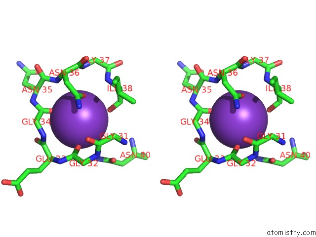
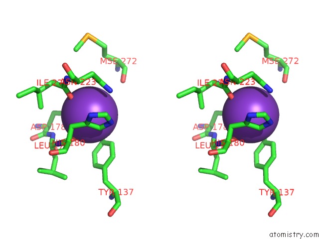
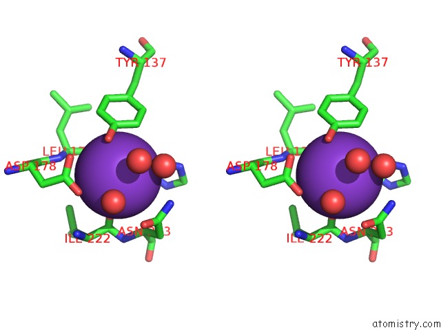
Potassium binding site 1 out of 6 in 4h5j

Go back to Potassium Binding Sites List in 4h5j
Potassium binding site 1 out of 6 in the Crystal Structure of the Guanine Nucleotide Exchange Factor SEC12 (P64 Form)


Mono view


Stereo pair view

A full contact list of Potassium with other atoms in the K binding site number 1 of Crystal Structure of the Guanine Nucleotide Exchange Factor SEC12 (P64 Form) within 5.0Å range:
probe atom residue distance (Å) B Occ
A:K401

b:30.1
occ:1.00
O A:GLY34 2.5 24.8 1.0
OD1 A:ASN36 2.7 28.1 1.0
O A:ASP68 2.8 38.4 1.0
O A:GLY32 2.8 32.1 1.0
O A:ILE38 3.0 31.7 1.0
CG A:ASN36 3.5 23.9 1.0
C A:GLY34 3.7 25.4 1.0
ND2 A:ASN36 3.8 16.7 1.0
C A:ASP68 3.8 32.4 1.0
C A:GLY32 4.0 23.4 1.0
N A:GLY34 4.0 21.5 1.0
C A:GLU33 4.1 25.2 1.0
CA A:ASP68 4.2 28.9 1.0
C A:ILE38 4.2 29.2 1.0
N A:ASN36 4.2 21.0 1.0
ND2 A:ASN40 4.2 31.2 1.0
CA A:GLY34 4.4 17.9 1.0
O A:GLU33 4.4 28.9 1.0
CB A:ASP68 4.4 32.0 1.0
CA A:GLU33 4.6 27.5 1.0
N A:ILE38 4.7 28.0 1.0
CB A:ASN36 4.7 25.2 1.0
N A:GLU33 4.7 25.1 1.0
N A:ASN35 4.7 22.6 1.0
N A:GLY32 4.9 25.2 1.0
CA A:ASN35 4.9 24.1 1.0
CA A:ILE38 4.9 26.8 1.0
CA A:ASN36 4.9 20.7 1.0
CG A:ASN40 4.9 29.4 1.0
N A:GLY37 5.0 23.0 1.0
OD1 A:ASP68 5.0 50.8 1.0
C A:GLY31 5.0 24.2 1.0

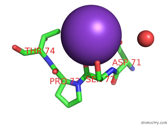
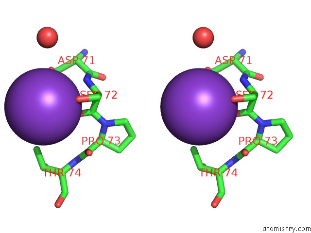
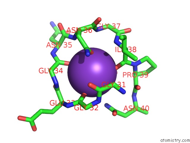
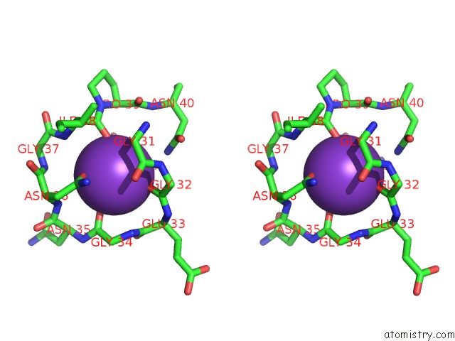
Potassium binding site 2 out of 6 in 4h5j

Go back to Potassium Binding Sites List in 4h5j
Potassium binding site 2 out of 6 in the Crystal Structure of the Guanine Nucleotide Exchange Factor SEC12 (P64 Form)


Mono view


Stereo pair view

A full contact list of Potassium with other atoms in the K binding site number 2 of Crystal Structure of the Guanine Nucleotide Exchange Factor SEC12 (P64 Form) within 5.0Å range:
probe atom residue distance (Å) B Occ
A:K402

b:41.3
occ:1.00
OG A:SER72 2.8 24.8 1.0
OD2 A:ASP71 2.9 27.4 1.0
O A:SER72 3.0 35.9 1.0
OD1 A:ASP71 3.0 31.2 1.0
CG A:ASP71 3.2 28.0 1.0
CG2 A:THR74 3.6 9.6 1.0
C A:SER72 3.8 30.0 1.0
N A:SER72 3.8 28.4 1.0
CB A:SER72 4.0 22.3 1.0
CA A:SER72 4.1 26.3 1.0
CB A:ASP71 4.5 22.4 1.0
O A:PRO73 4.7 24.8 1.0
C A:ASP71 4.8 24.3 1.0
C A:PRO73 4.9 24.4 1.0
O A:HOH505 4.9 28.3 1.0
N A:PRO73 5.0 27.3 1.0
CB A:THR74 5.0 23.1 1.0

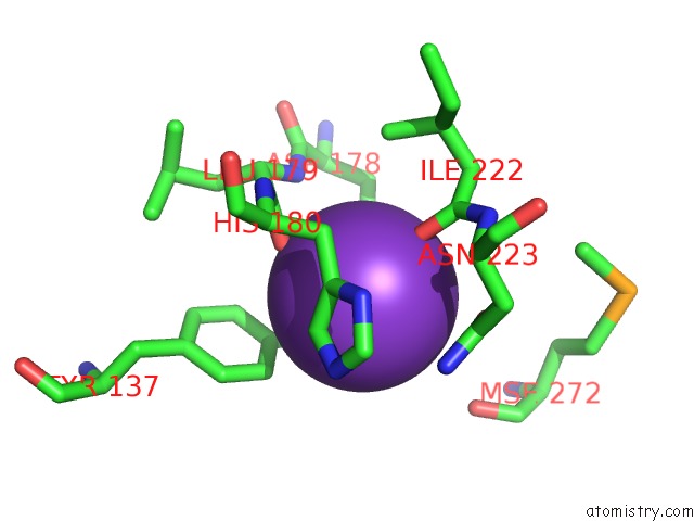
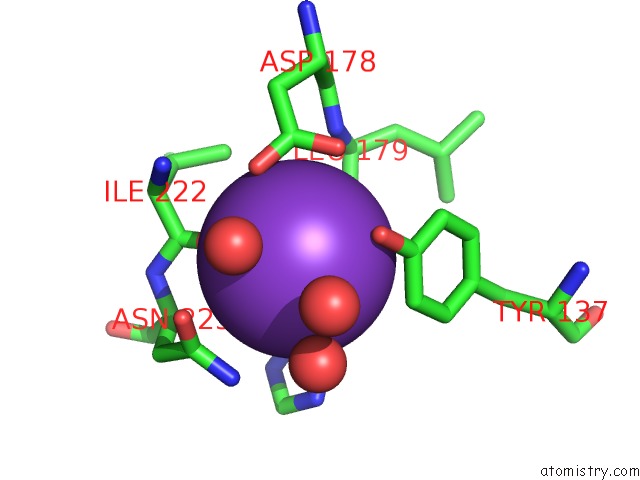
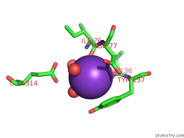
Potassium binding site 3 out of 6 in 4h5j

Go back to Potassium Binding Sites List in 4h5j
Potassium binding site 3 out of 6 in the Crystal Structure of the Guanine Nucleotide Exchange Factor SEC12 (P64 Form)


Mono view


Stereo pair view

A full contact list of Potassium with other atoms in the K binding site number 3 of Crystal Structure of the Guanine Nucleotide Exchange Factor SEC12 (P64 Form) within 5.0Å range:
probe atom residue distance (Å) B Occ
A:K403

b:41.9
occ:1.00
O A:ILE222 2.8 38.8 1.0
OH A:TYR137 2.8 23.7 1.0
O A:LEU179 2.8 39.7 1.0
OD1 A:ASP178 2.9 38.3 1.0
OD2 A:ASP178 3.2 34.1 1.0
CZ A:TYR137 3.3 22.9 1.0
CG A:ASP178 3.4 32.3 1.0
CE1 A:TYR137 3.6 25.1 1.0
OD1 A:ASN223 3.6 41.8 1.0
CD2 A:HIS180 3.7 35.1 1.0
ND2 A:ASN223 3.8 41.2 1.0
C A:LEU179 3.8 34.1 1.0
CG A:ASN223 3.9 42.5 1.0
C A:ILE222 3.9 34.2 1.0
CG A:HIS180 4.0 37.1 1.0
CB A:HIS180 4.0 34.8 1.0
CE2 A:TYR137 4.2 17.8 1.0
N A:LEU179 4.3 30.6 1.0
N A:ILE222 4.5 31.1 1.0
CA A:HIS180 4.6 34.7 1.0
CD1 A:TYR137 4.6 26.1 1.0
N A:HIS180 4.6 33.5 1.0
NE2 A:HIS180 4.7 32.9 1.0
CA A:LEU179 4.7 32.2 1.0
CB A:ASP178 4.7 28.5 1.0
CG2 A:ILE222 4.8 32.4 1.0
CA A:ILE222 4.8 32.8 1.0
N A:ASN223 4.8 32.0 1.0
O A:MSE272 4.9 35.2 1.0
CA A:ASN223 4.9 39.2 1.0

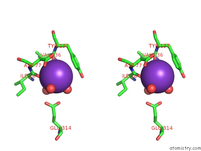
Potassium binding site 4 out of 6 in 4h5j

Go back to Potassium Binding Sites List in 4h5j
Potassium binding site 4 out of 6 in the Crystal Structure of the Guanine Nucleotide Exchange Factor SEC12 (P64 Form)


Mono view


Stereo pair view

A full contact list of Potassium with other atoms in the K binding site number 4 of Crystal Structure of the Guanine Nucleotide Exchange Factor SEC12 (P64 Form) within 5.0Å range:
probe atom residue distance (Å) B Occ
B:K401

b:22.0
occ:1.00
O B:GLY34 2.7 26.8 1.0
O B:GLY32 2.7 16.5 1.0
O B:ILE38 2.7 17.1 1.0
OD1 B:ASN36 2.8 23.1 1.0
O B:ASP68 2.9 24.1 1.0
CG B:ASN36 3.8 17.5 1.0
C B:ILE38 3.9 18.0 1.0
C B:GLY34 3.9 23.7 1.0
C B:GLY32 3.9 18.5 1.0
C B:ASP68 4.0 14.8 1.0
ND2 B:ASN40 4.1 21.2 1.0
N B:ASN36 4.3 28.2 1.0
N B:GLY34 4.3 22.0 1.0
C B:GLU33 4.3 20.6 1.0
N B:ILE38 4.3 16.3 1.0
CA B:ASP68 4.4 13.3 1.0
ND2 B:ASN36 4.4 13.6 1.0
C B:GLY31 4.4 14.7 1.0
N B:GLY32 4.5 14.5 1.0
O B:GLU33 4.5 22.9 1.0
CB B:ASP68 4.6 17.5 1.0
CA B:ILE38 4.6 17.8 1.0
CA B:GLY34 4.6 15.9 1.0
CA B:GLU33 4.7 17.7 1.0
O B:GLY31 4.7 22.7 1.0
N B:GLU33 4.7 17.7 1.0
N B:GLY37 4.8 14.8 1.0
CA B:GLY31 4.8 17.8 1.0
N B:PRO39 4.8 17.6 1.0
CA B:GLY32 4.8 7.3 1.0
N B:ASN35 4.9 27.8 1.0
CB B:ASN36 4.9 9.5 1.0
CA B:PRO39 5.0 14.9 1.0
CA B:ASN35 5.0 27.0 1.0
CG B:ASN40 5.0 21.3 1.0
CA B:ASN36 5.0 11.0 1.0

Potassium binding site 5 out of 6 in 4h5j

Go back to Potassium Binding Sites List in 4h5j
Potassium binding site 5 out of 6 in the Crystal Structure of the Guanine Nucleotide Exchange Factor SEC12 (P64 Form)


Mono view


Stereo pair view

A full contact list of Potassium with other atoms in the K binding site number 5 of Crystal Structure of the Guanine Nucleotide Exchange Factor SEC12 (P64 Form) within 5.0Å range:
probe atom residue distance (Å) B Occ
B:K402

b:25.0
occ:1.00
O B:ILE222 2.6 19.0 1.0
O B:LEU179 2.9 25.4 1.0
OD1 B:ASP178 2.9 16.5 1.0
OH B:TYR137 3.0 31.8 1.0
O B:HOH545 3.1 22.8 1.0
CZ B:TYR137 3.3 23.6 1.0
OD2 B:ASP178 3.4 21.8 1.0
CG B:ASP178 3.4 18.3 1.0
CE1 B:TYR137 3.5 22.2 1.0
CD2 B:HIS180 3.6 21.1 1.0
OD1 B:ASN223 3.6 32.8 1.0
O B:HOH555 3.6 35.5 1.0
ND2 B:ASN223 3.7 32.6 1.0
CG B:ASN223 3.8 32.5 1.0
C B:ILE222 3.8 17.3 1.0
C B:LEU179 3.8 22.6 1.0
CB B:HIS180 3.8 18.9 1.0
CG B:HIS180 3.9 20.4 1.0
CE2 B:TYR137 4.2 16.6 1.0
O B:HOH505 4.3 19.1 1.0
N B:LEU179 4.3 21.8 1.0
CA B:HIS180 4.4 22.7 1.0
CD1 B:TYR137 4.4 13.4 1.0
N B:HIS180 4.5 21.0 1.0
N B:ILE222 4.5 14.4 1.0
NE2 B:HIS180 4.6 9.2 1.0
N B:ASN223 4.6 15.9 1.0
CA B:ASN223 4.7 15.2 1.0
CG2 B:ILE222 4.7 21.2 1.0
CA B:LEU179 4.7 23.2 1.0
CA B:ILE222 4.7 16.2 1.0
CB B:ASP178 4.8 9.2 1.0
CB B:ASN223 4.8 21.8 1.0
ND1 B:HIS180 4.9 20.9 1.0
CD2 B:TYR137 5.0 18.7 1.0

Potassium binding site 6 out of 6 in 4h5j

Go back to Potassium Binding Sites List in 4h5j
Potassium binding site 6 out of 6 in the Crystal Structure of the Guanine Nucleotide Exchange Factor SEC12 (P64 Form)


Mono view


Stereo pair view

A full contact list of Potassium with other atoms in the K binding site number 6 of Crystal Structure of the Guanine Nucleotide Exchange Factor SEC12 (P64 Form) within 5.0Å range:
probe atom residue distance (Å) B Occ
B:K403

b:38.9
occ:1.00
O B:ILE76 2.7 13.1 1.0
O B:VAL136 2.7 24.2 1.0
O B:HOH553 2.7 30.3 1.0
O B:HOH538 2.9 21.3 1.0
O B:HOH510 3.3 15.1 1.0
O B:HOH554 3.3 37.4 1.0
CE1 B:TYR137 3.8 22.2 1.0
C B:ILE76 3.8 17.3 1.0
CZ B:TYR137 3.9 23.6 1.0
C B:VAL136 3.9 24.4 1.0
CD1 B:TYR137 4.1 13.4 1.0
CE2 B:TYR137 4.2 16.6 1.0
CB B:ASP77 4.3 18.8 1.0
OH B:TYR137 4.3 31.8 1.0
OE1 B:GLU314 4.3 37.1 1.0
CG B:ASP77 4.4 22.7 1.0
CG B:TYR137 4.4 15.6 1.0
N B:ILE76 4.5 11.8 1.0
N B:VAL136 4.5 20.5 1.0
CD2 B:TYR137 4.5 18.7 1.0
N B:ASP77 4.6 17.0 1.0
OE2 B:GLU314 4.6 35.1 1.0
CA B:VAL136 4.6 21.3 1.0
CA B:ASP77 4.7 14.5 1.0
OD1 B:ASP77 4.7 16.9 1.0
CA B:ILE76 4.8 13.8 1.0
OD2 B:ASP77 4.8 23.9 1.0
N B:TYR137 4.9 26.3 1.0
CB B:VAL136 4.9 18.8 1.0
CD B:GLU314 4.9 32.5 1.0
CA B:TYR137 5.0 21.0 1.0

Reference:

C.Mcmahon, S.M.Studer, C.Clendinen, G.P.Dann, P.D.Jeffrey, F.M.Hughson. The Structure of SEC12 Implicates Potassium Ion Coordination in SAR1 Activation. J.Biol.Chem. V. 287 43599 2012.
ISSN: ISSN 0021-9258
PubMed: 23109340
DOI: 10.1074/JBC.M112.420141
Page generated: Sun Dec 13 23:31:11 2020

Last articles

Zn in 8WB0
Zn in 8WAX
Zn in 8WAU
Zn in 8WAZ
Zn in 8WAY
Zn in 8WAV
Zn in 8WAW
Zn in 8WAT
Zn in 8W7M
Zn in 8WD3
© Copyright 2008-2020 by atomistry.com
Home   |    Site Map   |    Copyright   |    Contact us   |    Privacy