Potassium »
PDB 4eei-4gx0 »
4glh »
Potassium in PDB 4glh: Dna Dodecamer Containing 5-Hydroxymethyl Cytosine
Protein crystallography data
The structure of Dna Dodecamer Containing 5-Hydroxymethyl Cytosine, PDB code: 4glh
was solved by
B.Spingler,
D.Renciuk,
M.Vorlickova,
with X-Ray Crystallography technique. A brief refinement statistics is given in the table below:
| Resolution Low / High (Å) | 41.78 / 1.66 |
| Space group | P 21 21 21 |
| Cell size a, b, c (Å), α, β, γ (°) | 25.637, 41.767, 63.997, 90.00, 90.00, 90.00 |
| R / Rfree (%) | 23.7 / 27.6 |
Potassium Binding Sites:
The binding sites of Potassium atom in the Dna Dodecamer Containing 5-Hydroxymethyl Cytosine
(pdb code 4glh). This binding sites where shown within
5.0 Angstroms radius around Potassium atom.
In total 2 binding sites of Potassium where determined in the Dna Dodecamer Containing 5-Hydroxymethyl Cytosine, PDB code: 4glh:
Jump to Potassium binding site number: 1; 2;
In total 2 binding sites of Potassium where determined in the Dna Dodecamer Containing 5-Hydroxymethyl Cytosine, PDB code: 4glh:
Jump to Potassium binding site number: 1; 2;
Potassium binding site 1 out of 2 in 4glh
Go back to
Potassium binding site 1 out
of 2 in the Dna Dodecamer Containing 5-Hydroxymethyl Cytosine
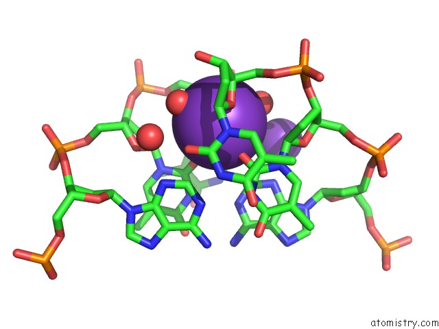
Mono view
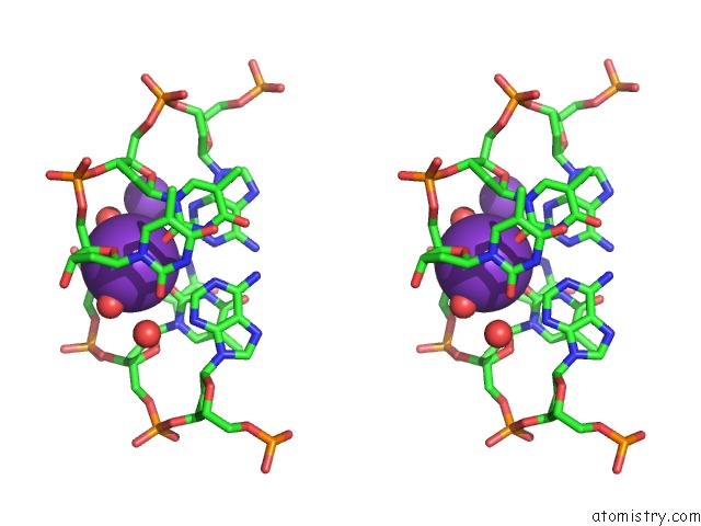
Stereo pair view
Mono view
Stereo pair view
A full contact list of Potassium with other atoms in the K binding
site number 1 of Dna Dodecamer Containing 5-Hydroxymethyl Cytosine within 5.0Å range:
|
Potassium binding site 2 out of 2 in 4glh
Go back to
Potassium binding site 2 out
of 2 in the Dna Dodecamer Containing 5-Hydroxymethyl Cytosine
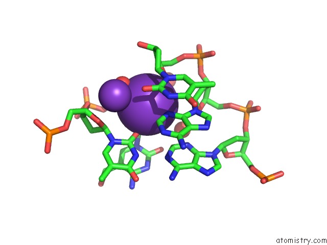
Mono view
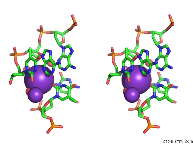
Stereo pair view
Mono view
Stereo pair view
A full contact list of Potassium with other atoms in the K binding
site number 2 of Dna Dodecamer Containing 5-Hydroxymethyl Cytosine within 5.0Å range:
|
Reference:
D.Renciuk,
O.Blacque,
M.Vorlickova,
B.Spingler.
Crystal Structures of B-Dna Dodecamer Containing the Epigenetic Modifications 5-Hydroxymethylcytosine or 5-Methylcytosine. Nucleic Acids Res. V. 41 9891 2013.
ISSN: ISSN 0305-1048
PubMed: 23963698
DOI: 10.1093/NAR/GKT738
Page generated: Mon Aug 12 10:49:48 2024
ISSN: ISSN 0305-1048
PubMed: 23963698
DOI: 10.1093/NAR/GKT738
Last articles
Zn in 9J0NZn in 9J0O
Zn in 9J0P
Zn in 9FJX
Zn in 9EKB
Zn in 9C0F
Zn in 9CAH
Zn in 9CH0
Zn in 9CH3
Zn in 9CH1