Potassium »
PDB 4eei-4gx0 »
4fb0 »
Potassium in PDB 4fb0: Structure of Oceanobacillus Iheyensis Group II Intron C377G Mutant in A Ligand-Free State in the Presence of K+ and MG2+
Protein crystallography data
The structure of Structure of Oceanobacillus Iheyensis Group II Intron C377G Mutant in A Ligand-Free State in the Presence of K+ and MG2+, PDB code: 4fb0
was solved by
M.Marcia,
A.M.Pyle,
with X-Ray Crystallography technique. A brief refinement statistics is given in the table below:
| Resolution Low / High (Å) | 49.16 / 3.22 |
| Space group | P 21 21 21 |
| Cell size a, b, c (Å), α, β, γ (°) | 89.188, 95.444, 224.706, 90.00, 90.00, 90.00 |
| R / Rfree (%) | 18.4 / 23 |
Other elements in 4fb0:
The structure of Structure of Oceanobacillus Iheyensis Group II Intron C377G Mutant in A Ligand-Free State in the Presence of K+ and MG2+ also contains other interesting chemical elements:
| Magnesium | (Mg) | 25 atoms |
Potassium Binding Sites:
Pages:
>>> Page 1 <<< Page 2, Binding sites: 11 - 18;Binding sites:
The binding sites of Potassium atom in the Structure of Oceanobacillus Iheyensis Group II Intron C377G Mutant in A Ligand-Free State in the Presence of K+ and MG2+ (pdb code 4fb0). This binding sites where shown within 5.0 Angstroms radius around Potassium atom.In total 18 binding sites of Potassium where determined in the Structure of Oceanobacillus Iheyensis Group II Intron C377G Mutant in A Ligand-Free State in the Presence of K+ and MG2+, PDB code: 4fb0:
Jump to Potassium binding site number: 1; 2; 3; 4; 5; 6; 7; 8; 9; 10;
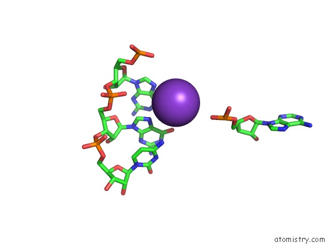
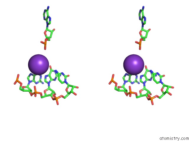
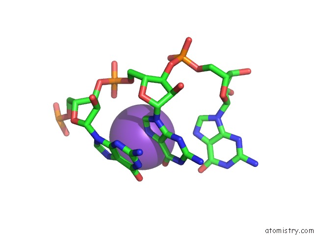
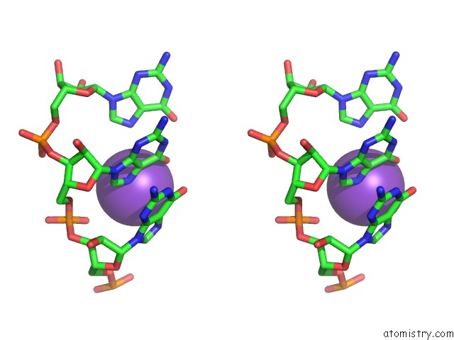
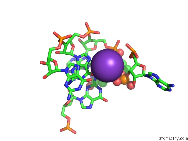
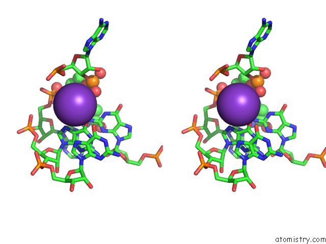
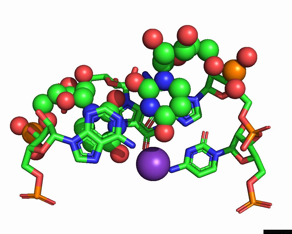
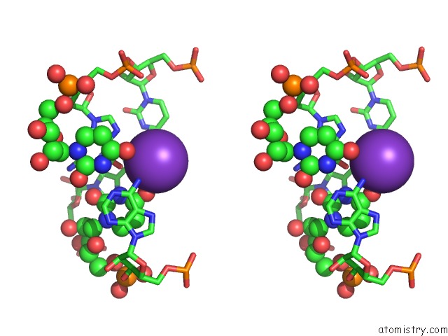
Potassium binding site 1 out of 18 in 4fb0
Go back to
Potassium binding site 1 out
of 18 in the Structure of Oceanobacillus Iheyensis Group II Intron C377G Mutant in A Ligand-Free State in the Presence of K+ and MG2+
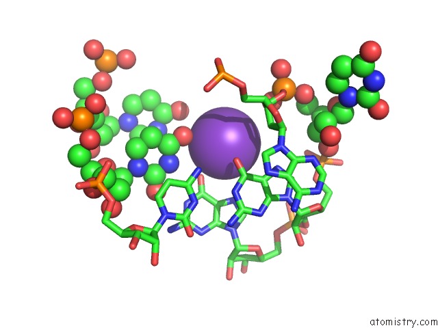
Mono view
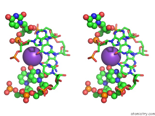
Stereo pair view
Mono view
Stereo pair view
A full contact list of Potassium with other atoms in the K binding
site number 1 of Structure of Oceanobacillus Iheyensis Group II Intron C377G Mutant in A Ligand-Free State in the Presence of K+ and MG2+ within 5.0Å range:
|
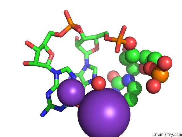
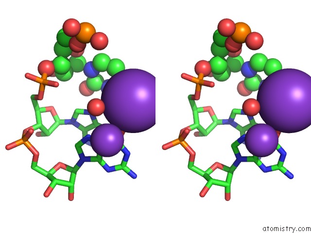
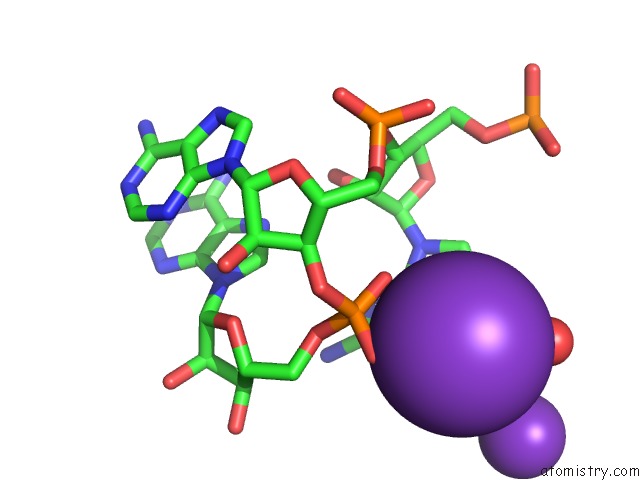
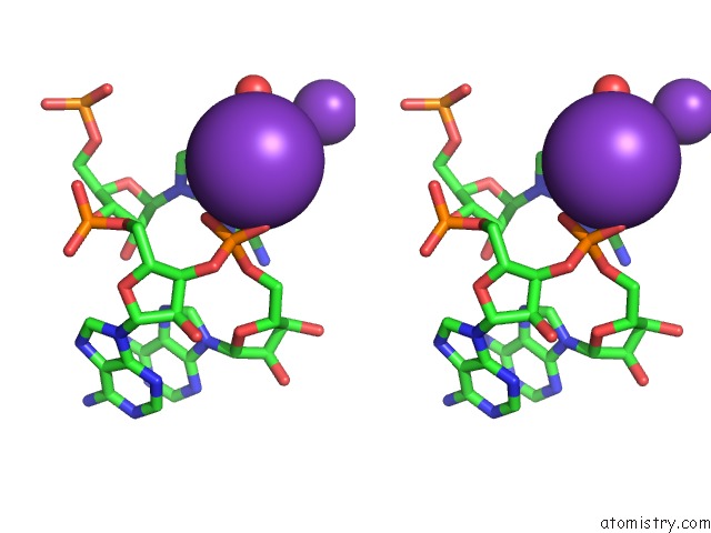
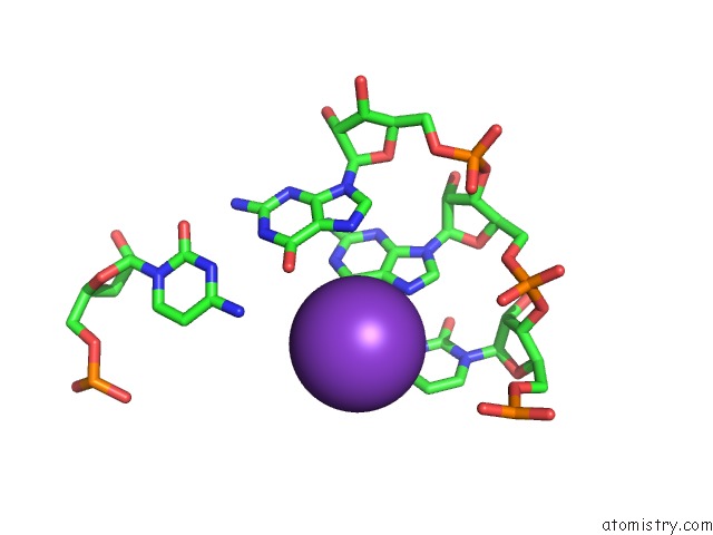
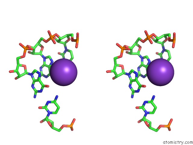
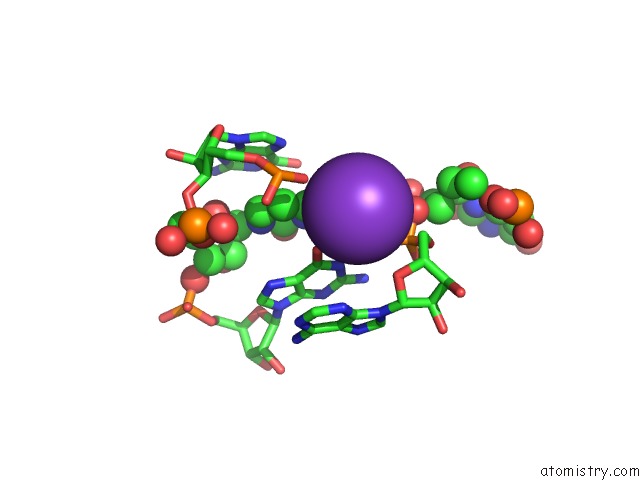
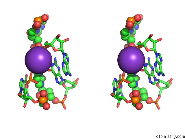
Potassium binding site 2 out of 18 in 4fb0
Go back to
Potassium binding site 2 out
of 18 in the Structure of Oceanobacillus Iheyensis Group II Intron C377G Mutant in A Ligand-Free State in the Presence of K+ and MG2+
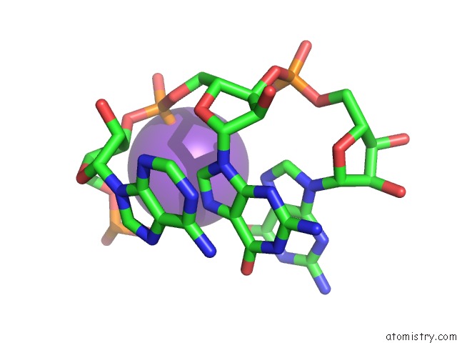
Mono view
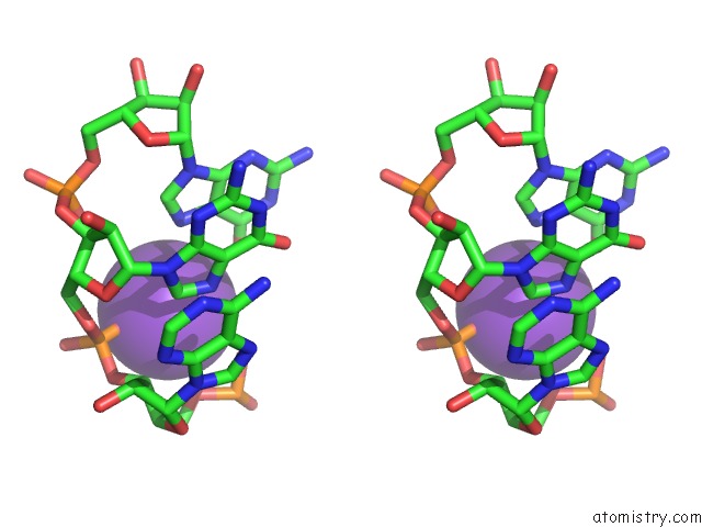
Stereo pair view
Mono view
Stereo pair view
A full contact list of Potassium with other atoms in the K binding
site number 2 of Structure of Oceanobacillus Iheyensis Group II Intron C377G Mutant in A Ligand-Free State in the Presence of K+ and MG2+ within 5.0Å range:
|
Potassium binding site 3 out of 18 in 4fb0
Go back to
Potassium binding site 3 out
of 18 in the Structure of Oceanobacillus Iheyensis Group II Intron C377G Mutant in A Ligand-Free State in the Presence of K+ and MG2+
Mono view
Stereo pair view
Mono view
Stereo pair view
A full contact list of Potassium with other atoms in the K binding
site number 3 of Structure of Oceanobacillus Iheyensis Group II Intron C377G Mutant in A Ligand-Free State in the Presence of K+ and MG2+ within 5.0Å range:
|
Potassium binding site 4 out of 18 in 4fb0
Go back to
Potassium binding site 4 out
of 18 in the Structure of Oceanobacillus Iheyensis Group II Intron C377G Mutant in A Ligand-Free State in the Presence of K+ and MG2+
Mono view
Stereo pair view
Mono view
Stereo pair view
A full contact list of Potassium with other atoms in the K binding
site number 4 of Structure of Oceanobacillus Iheyensis Group II Intron C377G Mutant in A Ligand-Free State in the Presence of K+ and MG2+ within 5.0Å range:
|
Potassium binding site 5 out of 18 in 4fb0
Go back to
Potassium binding site 5 out
of 18 in the Structure of Oceanobacillus Iheyensis Group II Intron C377G Mutant in A Ligand-Free State in the Presence of K+ and MG2+
Mono view
Stereo pair view
Mono view
Stereo pair view
A full contact list of Potassium with other atoms in the K binding
site number 5 of Structure of Oceanobacillus Iheyensis Group II Intron C377G Mutant in A Ligand-Free State in the Presence of K+ and MG2+ within 5.0Å range:
|
Potassium binding site 6 out of 18 in 4fb0
Go back to
Potassium binding site 6 out
of 18 in the Structure of Oceanobacillus Iheyensis Group II Intron C377G Mutant in A Ligand-Free State in the Presence of K+ and MG2+
Mono view
Stereo pair view
Mono view
Stereo pair view
A full contact list of Potassium with other atoms in the K binding
site number 6 of Structure of Oceanobacillus Iheyensis Group II Intron C377G Mutant in A Ligand-Free State in the Presence of K+ and MG2+ within 5.0Å range:
|
Potassium binding site 7 out of 18 in 4fb0
Go back to
Potassium binding site 7 out
of 18 in the Structure of Oceanobacillus Iheyensis Group II Intron C377G Mutant in A Ligand-Free State in the Presence of K+ and MG2+
Mono view
Stereo pair view
Mono view
Stereo pair view
A full contact list of Potassium with other atoms in the K binding
site number 7 of Structure of Oceanobacillus Iheyensis Group II Intron C377G Mutant in A Ligand-Free State in the Presence of K+ and MG2+ within 5.0Å range:
|
Potassium binding site 8 out of 18 in 4fb0
Go back to
Potassium binding site 8 out
of 18 in the Structure of Oceanobacillus Iheyensis Group II Intron C377G Mutant in A Ligand-Free State in the Presence of K+ and MG2+
Mono view
Stereo pair view
Mono view
Stereo pair view
A full contact list of Potassium with other atoms in the K binding
site number 8 of Structure of Oceanobacillus Iheyensis Group II Intron C377G Mutant in A Ligand-Free State in the Presence of K+ and MG2+ within 5.0Å range:
|
Potassium binding site 9 out of 18 in 4fb0
Go back to
Potassium binding site 9 out
of 18 in the Structure of Oceanobacillus Iheyensis Group II Intron C377G Mutant in A Ligand-Free State in the Presence of K+ and MG2+
Mono view
Stereo pair view
Mono view
Stereo pair view
A full contact list of Potassium with other atoms in the K binding
site number 9 of Structure of Oceanobacillus Iheyensis Group II Intron C377G Mutant in A Ligand-Free State in the Presence of K+ and MG2+ within 5.0Å range:
|
Potassium binding site 10 out of 18 in 4fb0
Go back to
Potassium binding site 10 out
of 18 in the Structure of Oceanobacillus Iheyensis Group II Intron C377G Mutant in A Ligand-Free State in the Presence of K+ and MG2+
Mono view
Stereo pair view
Mono view
Stereo pair view
A full contact list of Potassium with other atoms in the K binding
site number 10 of Structure of Oceanobacillus Iheyensis Group II Intron C377G Mutant in A Ligand-Free State in the Presence of K+ and MG2+ within 5.0Å range:
|
Reference:
M.Marcia,
A.M.Pyle.
Visualizing Group II Intron Catalysis Through the Stages of Splicing. Cell(Cambridge,Mass.) V. 151 497 2012.
ISSN: ISSN 0092-8674
PubMed: 23101623
DOI: 10.1016/J.CELL.2012.09.033
Page generated: Mon Aug 12 10:42:09 2024
ISSN: ISSN 0092-8674
PubMed: 23101623
DOI: 10.1016/J.CELL.2012.09.033
Last articles
Zn in 9J0NZn in 9J0O
Zn in 9J0P
Zn in 9FJX
Zn in 9EKB
Zn in 9C0F
Zn in 9CAH
Zn in 9CH0
Zn in 9CH3
Zn in 9CH1