Potassium »
PDB 4eei-4gx0 »
4eyv »
Potassium in PDB 4eyv: Crystal Structure of Cyclophilin A Like Protein From Piriformospora Indica
Enzymatic activity of Crystal Structure of Cyclophilin A Like Protein From Piriformospora Indica
All present enzymatic activity of Crystal Structure of Cyclophilin A Like Protein From Piriformospora Indica:
5.2.1.8;
5.2.1.8;
Protein crystallography data
The structure of Crystal Structure of Cyclophilin A Like Protein From Piriformospora Indica, PDB code: 4eyv
was solved by
H.Bhatt,
R.K.Pal,
N.Tuteja,
N.S.Bhavesh,
with X-Ray Crystallography technique. A brief refinement statistics is given in the table below:
| Resolution Low / High (Å) | 27.03 / 1.97 |
| Space group | C 2 2 21 |
| Cell size a, b, c (Å), α, β, γ (°) | 121.252, 144.108, 110.735, 90.00, 90.00, 90.00 |
| R / Rfree (%) | 15.9 / 18.3 |
Other elements in 4eyv:
The structure of Crystal Structure of Cyclophilin A Like Protein From Piriformospora Indica also contains other interesting chemical elements:
| Sodium | (Na) | 10 atoms |
Potassium Binding Sites:
The binding sites of Potassium atom in the Crystal Structure of Cyclophilin A Like Protein From Piriformospora Indica
(pdb code 4eyv). This binding sites where shown within
5.0 Angstroms radius around Potassium atom.
In total 4 binding sites of Potassium where determined in the Crystal Structure of Cyclophilin A Like Protein From Piriformospora Indica, PDB code: 4eyv:
Jump to Potassium binding site number: 1; 2; 3; 4;
In total 4 binding sites of Potassium where determined in the Crystal Structure of Cyclophilin A Like Protein From Piriformospora Indica, PDB code: 4eyv:
Jump to Potassium binding site number: 1; 2; 3; 4;
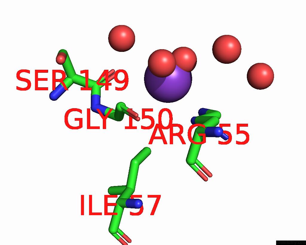
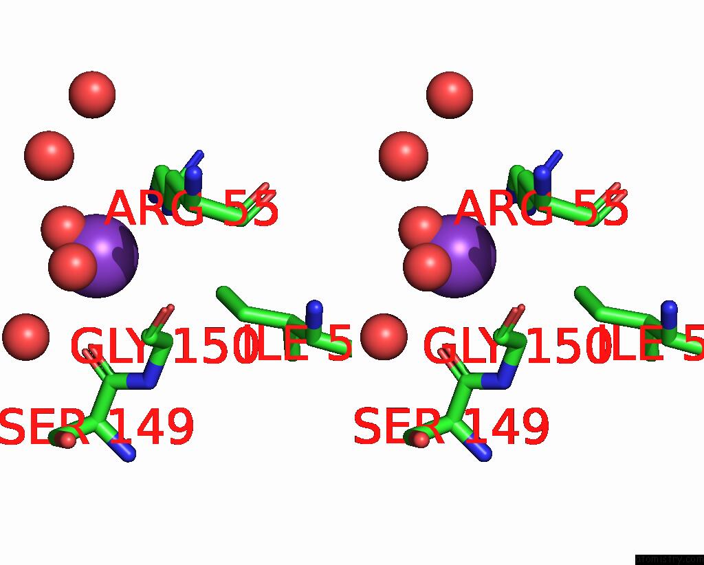
Potassium binding site 1 out of 4 in 4eyv
Go back to
Potassium binding site 1 out
of 4 in the Crystal Structure of Cyclophilin A Like Protein From Piriformospora Indica
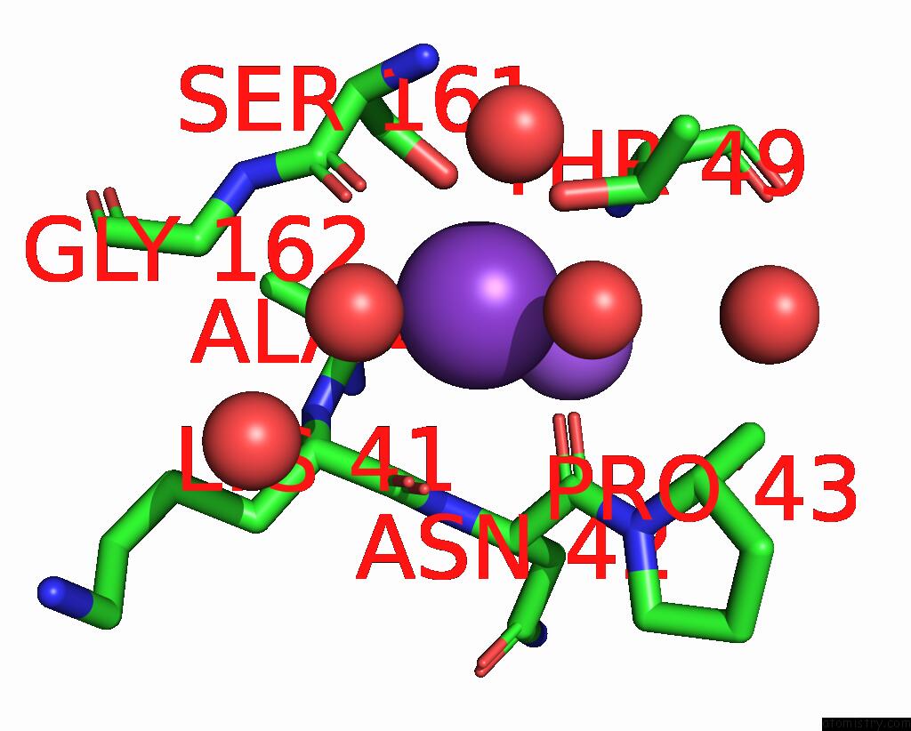
Mono view
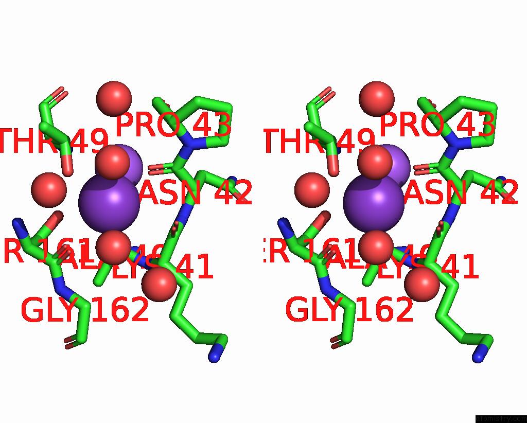
Stereo pair view
Mono view
Stereo pair view
A full contact list of Potassium with other atoms in the K binding
site number 1 of Crystal Structure of Cyclophilin A Like Protein From Piriformospora Indica within 5.0Å range:
|
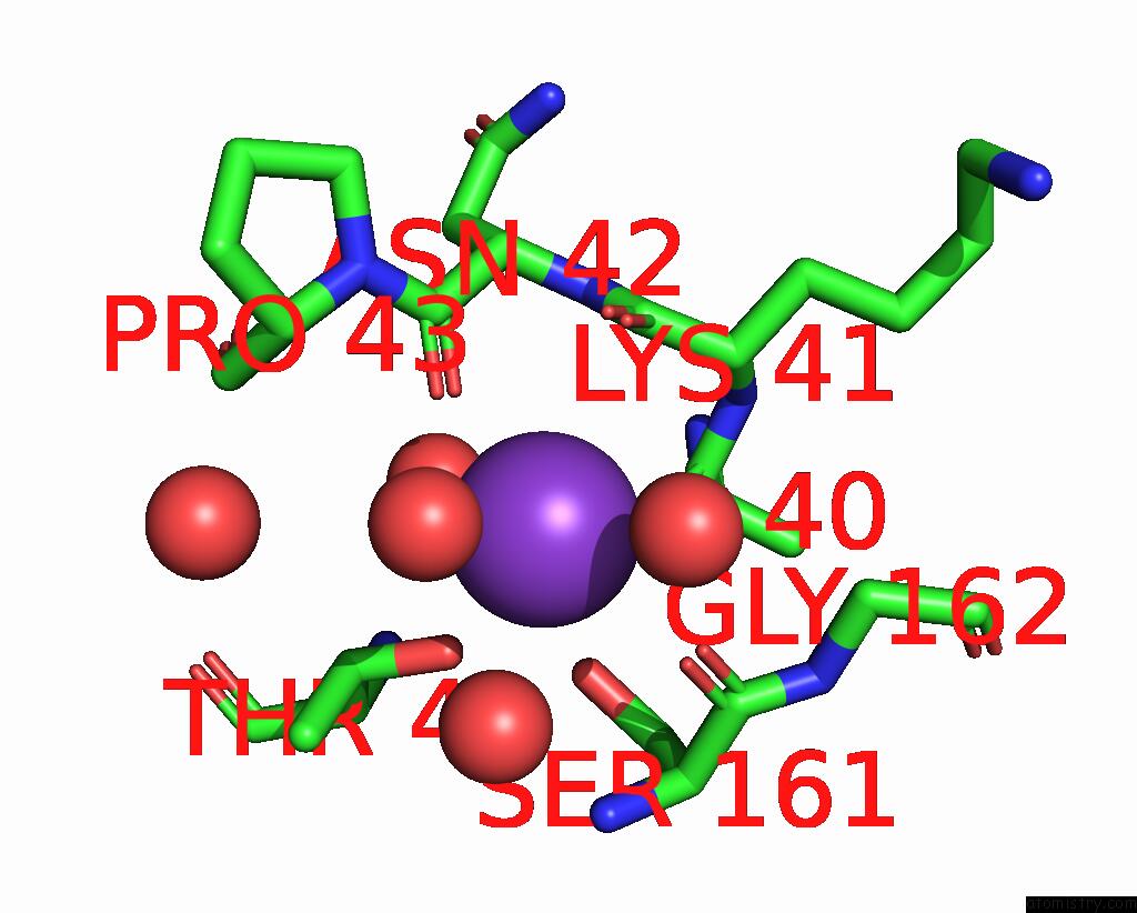
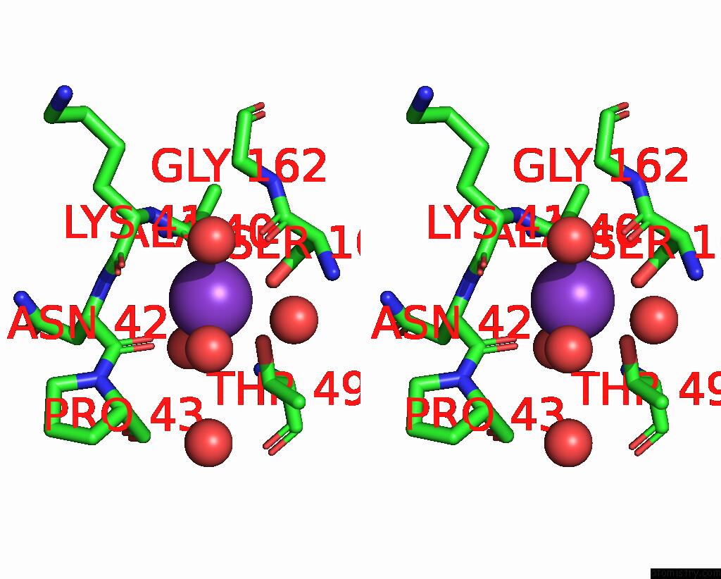
Potassium binding site 2 out of 4 in 4eyv
Go back to
Potassium binding site 2 out
of 4 in the Crystal Structure of Cyclophilin A Like Protein From Piriformospora Indica
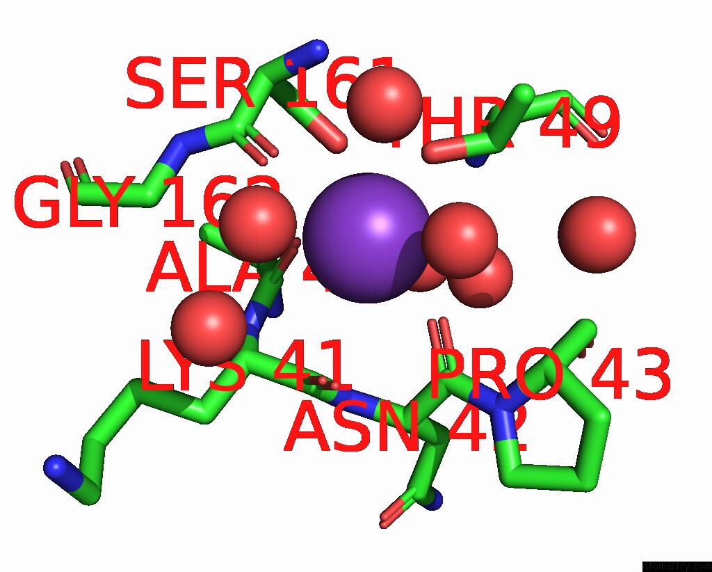
Mono view
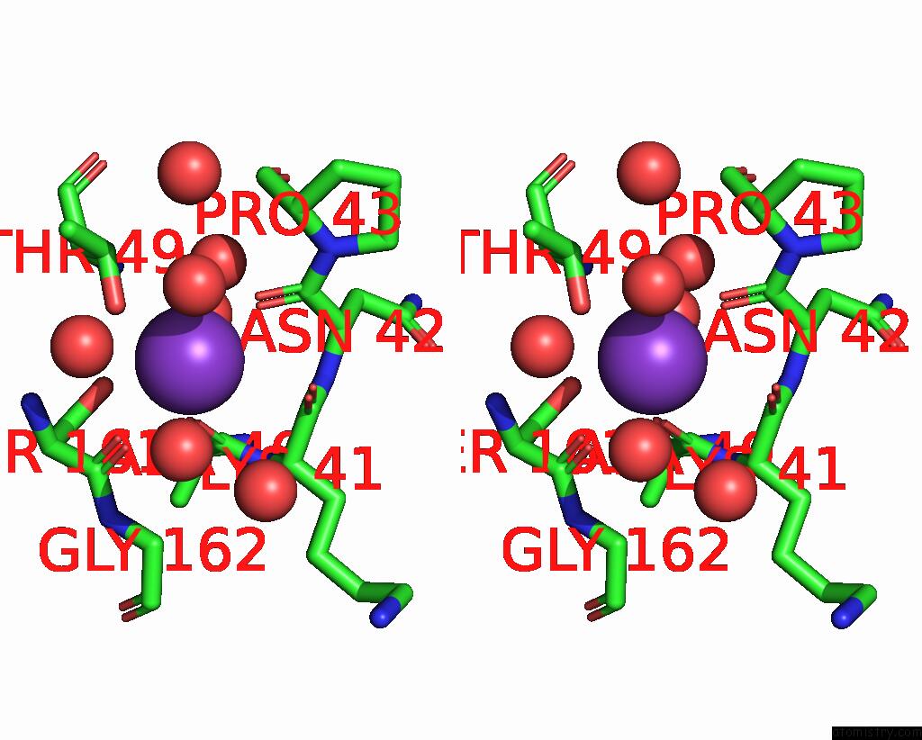
Stereo pair view
Mono view
Stereo pair view
A full contact list of Potassium with other atoms in the K binding
site number 2 of Crystal Structure of Cyclophilin A Like Protein From Piriformospora Indica within 5.0Å range:
|
Potassium binding site 3 out of 4 in 4eyv
Go back to
Potassium binding site 3 out
of 4 in the Crystal Structure of Cyclophilin A Like Protein From Piriformospora Indica
Mono view
Stereo pair view
Mono view
Stereo pair view
A full contact list of Potassium with other atoms in the K binding
site number 3 of Crystal Structure of Cyclophilin A Like Protein From Piriformospora Indica within 5.0Å range:
|
Potassium binding site 4 out of 4 in 4eyv
Go back to
Potassium binding site 4 out
of 4 in the Crystal Structure of Cyclophilin A Like Protein From Piriformospora Indica
Mono view
Stereo pair view
Mono view
Stereo pair view
A full contact list of Potassium with other atoms in the K binding
site number 4 of Crystal Structure of Cyclophilin A Like Protein From Piriformospora Indica within 5.0Å range:
|
Reference:
D.K.Trivedi,
H.Bhatt,
R.K.Pal,
R.Tuteja,
B.Garg,
A.K.Johri,
N.S.Bhavesh,
N.Tuteja.
Structure of Rna-Interacting Cyclophilin A-Like Protein From Piriformospora Indica That Provides Salinity-Stress Tolerance in Plants Sci Rep V. 3 3001 2013.
ISSN: ESSN 2045-2322
PubMed: 24141523
DOI: 10.1038/SREP03001
Page generated: Mon Aug 12 10:41:04 2024
ISSN: ESSN 2045-2322
PubMed: 24141523
DOI: 10.1038/SREP03001
Last articles
Zn in 9J0NZn in 9J0O
Zn in 9J0P
Zn in 9FJX
Zn in 9EKB
Zn in 9C0F
Zn in 9CAH
Zn in 9CH0
Zn in 9CH3
Zn in 9CH1