Potassium »
PDB 4eei-4gx0 »
4evy »
Potassium in PDB 4evy: Crystal Structure of Aminoglycoside Antibiotic 6'-N-Acetyltransferase Aac(6')-Ig From Acinetobacter Haemolyticus in Complex with Tobramycin
Enzymatic activity of Crystal Structure of Aminoglycoside Antibiotic 6'-N-Acetyltransferase Aac(6')-Ig From Acinetobacter Haemolyticus in Complex with Tobramycin
All present enzymatic activity of Crystal Structure of Aminoglycoside Antibiotic 6'-N-Acetyltransferase Aac(6')-Ig From Acinetobacter Haemolyticus in Complex with Tobramycin:
2.3.1.82;
2.3.1.82;
Protein crystallography data
The structure of Crystal Structure of Aminoglycoside Antibiotic 6'-N-Acetyltransferase Aac(6')-Ig From Acinetobacter Haemolyticus in Complex with Tobramycin, PDB code: 4evy
was solved by
P.J.Stogios,
E.Evdokimova,
G.Minasov,
V.Yim,
P.Courvalin,
A.Savchenko,
W.F.Anderson,
Center For Structural Genomics Of Infectious Diseases(Csgid),
with X-Ray Crystallography technique. A brief refinement statistics is given in the table below:
| Resolution Low / High (Å) | 28.97 / 1.77 |
| Space group | P 1 |
| Cell size a, b, c (Å), α, β, γ (°) | 39.723, 43.789, 46.282, 83.65, 87.49, 68.41 |
| R / Rfree (%) | 16.7 / 21.3 |
Other elements in 4evy:
The structure of Crystal Structure of Aminoglycoside Antibiotic 6'-N-Acetyltransferase Aac(6')-Ig From Acinetobacter Haemolyticus in Complex with Tobramycin also contains other interesting chemical elements:
| Chlorine | (Cl) | 3 atoms |
Potassium Binding Sites:
The binding sites of Potassium atom in the Crystal Structure of Aminoglycoside Antibiotic 6'-N-Acetyltransferase Aac(6')-Ig From Acinetobacter Haemolyticus in Complex with Tobramycin
(pdb code 4evy). This binding sites where shown within
5.0 Angstroms radius around Potassium atom.
In total 2 binding sites of Potassium where determined in the Crystal Structure of Aminoglycoside Antibiotic 6'-N-Acetyltransferase Aac(6')-Ig From Acinetobacter Haemolyticus in Complex with Tobramycin, PDB code: 4evy:
Jump to Potassium binding site number: 1; 2;
In total 2 binding sites of Potassium where determined in the Crystal Structure of Aminoglycoside Antibiotic 6'-N-Acetyltransferase Aac(6')-Ig From Acinetobacter Haemolyticus in Complex with Tobramycin, PDB code: 4evy:
Jump to Potassium binding site number: 1; 2;
Potassium binding site 1 out of 2 in 4evy
Go back to
Potassium binding site 1 out
of 2 in the Crystal Structure of Aminoglycoside Antibiotic 6'-N-Acetyltransferase Aac(6')-Ig From Acinetobacter Haemolyticus in Complex with Tobramycin
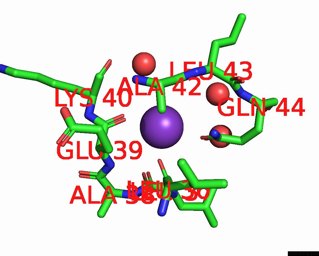
Mono view
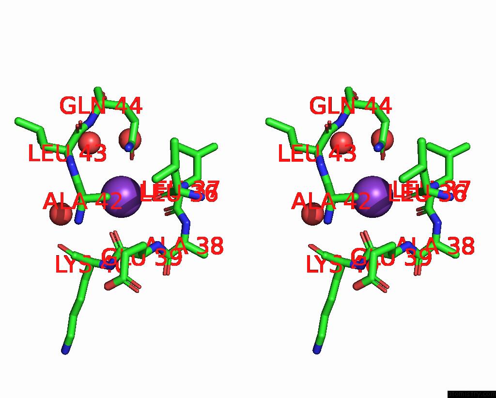
Stereo pair view
Mono view
Stereo pair view
A full contact list of Potassium with other atoms in the K binding
site number 1 of Crystal Structure of Aminoglycoside Antibiotic 6'-N-Acetyltransferase Aac(6')-Ig From Acinetobacter Haemolyticus in Complex with Tobramycin within 5.0Å range:
|
Potassium binding site 2 out of 2 in 4evy
Go back to
Potassium binding site 2 out
of 2 in the Crystal Structure of Aminoglycoside Antibiotic 6'-N-Acetyltransferase Aac(6')-Ig From Acinetobacter Haemolyticus in Complex with Tobramycin
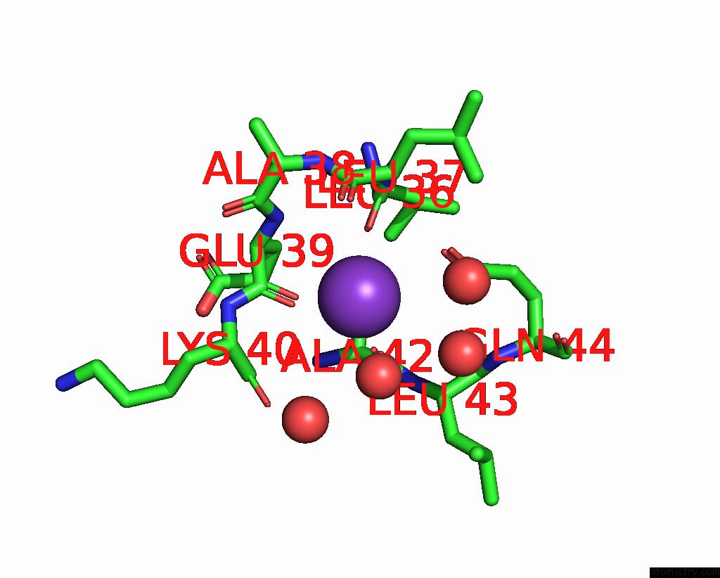
Mono view
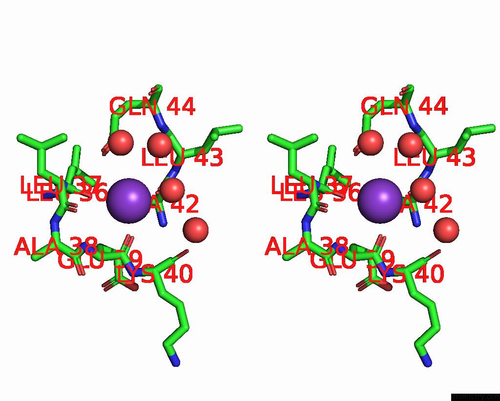
Stereo pair view
Mono view
Stereo pair view
A full contact list of Potassium with other atoms in the K binding
site number 2 of Crystal Structure of Aminoglycoside Antibiotic 6'-N-Acetyltransferase Aac(6')-Ig From Acinetobacter Haemolyticus in Complex with Tobramycin within 5.0Å range:
|
Reference:
P.J.Stogios,
M.L.Kuhn,
E.Evdokimova,
M.Law,
P.Courvalin,
A.Savchenko.
Structural and Biochemical Characterization of Acinetobacter Spp. Aminoglycoside Acetyltransferases Highlights Functional and Evolutionary Variation Among Antibiotic Resistance Enzymes. Acs Infect Dis. V. 3 132 2017.
ISSN: ESSN 2373-8227
PubMed: 27785912
DOI: 10.1021/ACSINFECDIS.6B00058
Page generated: Sat Aug 9 06:46:00 2025
ISSN: ESSN 2373-8227
PubMed: 27785912
DOI: 10.1021/ACSINFECDIS.6B00058
Last articles
Mg in 3GGIMg in 3GFT
Mg in 3GF0
Mg in 3GEB
Mg in 3GCM
Mg in 3GEI
Mg in 3GDX
Mg in 3GBJ
Mg in 3GDD
Mg in 3GAO