Potassium »
PDB 4eei-4gx0 »
4eou »
Potassium in PDB 4eou: Crystal Structure of E. Coli Dihydrodipicolinate Synthase with Pyruvate and Succinic Semi-Aldehyde Bound in Active Site
Enzymatic activity of Crystal Structure of E. Coli Dihydrodipicolinate Synthase with Pyruvate and Succinic Semi-Aldehyde Bound in Active Site
All present enzymatic activity of Crystal Structure of E. Coli Dihydrodipicolinate Synthase with Pyruvate and Succinic Semi-Aldehyde Bound in Active Site:
4.2.1.52;
4.2.1.52;
Protein crystallography data
The structure of Crystal Structure of E. Coli Dihydrodipicolinate Synthase with Pyruvate and Succinic Semi-Aldehyde Bound in Active Site, PDB code: 4eou
was solved by
B.A.Boughton,
R.C.J.Dobson,
C.A.Hutton,
with X-Ray Crystallography technique. A brief refinement statistics is given in the table below:
| Resolution Low / High (Å) | 29.10 / 2.30 |
| Space group | P 31 2 1 |
| Cell size a, b, c (Å), α, β, γ (°) | 121.172, 121.172, 109.732, 90.00, 90.00, 120.00 |
| R / Rfree (%) | 14.3 / 19.8 |
Potassium Binding Sites:
The binding sites of Potassium atom in the Crystal Structure of E. Coli Dihydrodipicolinate Synthase with Pyruvate and Succinic Semi-Aldehyde Bound in Active Site
(pdb code 4eou). This binding sites where shown within
5.0 Angstroms radius around Potassium atom.
In total 4 binding sites of Potassium where determined in the Crystal Structure of E. Coli Dihydrodipicolinate Synthase with Pyruvate and Succinic Semi-Aldehyde Bound in Active Site, PDB code: 4eou:
Jump to Potassium binding site number: 1; 2; 3; 4;
In total 4 binding sites of Potassium where determined in the Crystal Structure of E. Coli Dihydrodipicolinate Synthase with Pyruvate and Succinic Semi-Aldehyde Bound in Active Site, PDB code: 4eou:
Jump to Potassium binding site number: 1; 2; 3; 4;
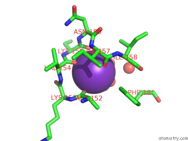
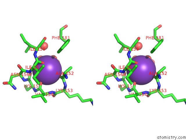
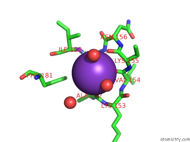
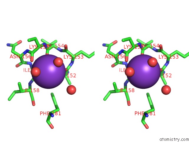
Potassium binding site 1 out of 4 in 4eou
Go back to
Potassium binding site 1 out
of 4 in the Crystal Structure of E. Coli Dihydrodipicolinate Synthase with Pyruvate and Succinic Semi-Aldehyde Bound in Active Site
Mono view
Stereo pair view
Mono view
Stereo pair view
A full contact list of Potassium with other atoms in the K binding
site number 1 of Crystal Structure of E. Coli Dihydrodipicolinate Synthase with Pyruvate and Succinic Semi-Aldehyde Bound in Active Site within 5.0Å range:
|
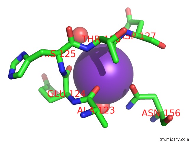
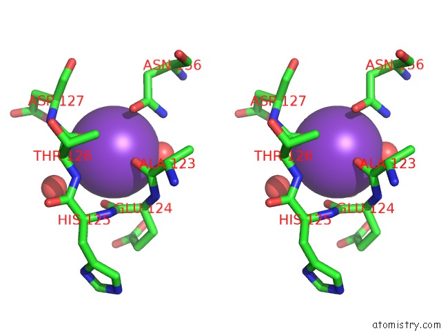
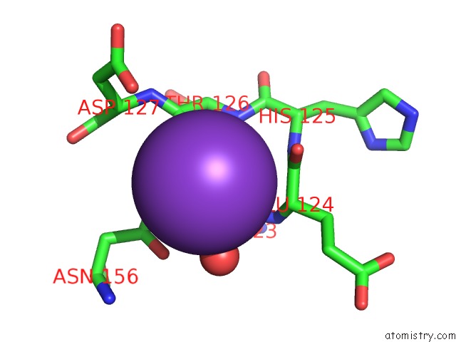
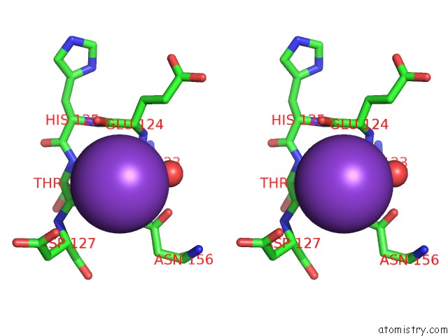
Potassium binding site 2 out of 4 in 4eou
Go back to
Potassium binding site 2 out
of 4 in the Crystal Structure of E. Coli Dihydrodipicolinate Synthase with Pyruvate and Succinic Semi-Aldehyde Bound in Active Site
Mono view
Stereo pair view
Mono view
Stereo pair view
A full contact list of Potassium with other atoms in the K binding
site number 2 of Crystal Structure of E. Coli Dihydrodipicolinate Synthase with Pyruvate and Succinic Semi-Aldehyde Bound in Active Site within 5.0Å range:
|
Potassium binding site 3 out of 4 in 4eou
Go back to
Potassium binding site 3 out
of 4 in the Crystal Structure of E. Coli Dihydrodipicolinate Synthase with Pyruvate and Succinic Semi-Aldehyde Bound in Active Site
Mono view
Stereo pair view
Mono view
Stereo pair view
A full contact list of Potassium with other atoms in the K binding
site number 3 of Crystal Structure of E. Coli Dihydrodipicolinate Synthase with Pyruvate and Succinic Semi-Aldehyde Bound in Active Site within 5.0Å range:
|
Potassium binding site 4 out of 4 in 4eou
Go back to
Potassium binding site 4 out
of 4 in the Crystal Structure of E. Coli Dihydrodipicolinate Synthase with Pyruvate and Succinic Semi-Aldehyde Bound in Active Site
Mono view
Stereo pair view
Mono view
Stereo pair view
A full contact list of Potassium with other atoms in the K binding
site number 4 of Crystal Structure of E. Coli Dihydrodipicolinate Synthase with Pyruvate and Succinic Semi-Aldehyde Bound in Active Site within 5.0Å range:
|
Reference:
B.A.Boughton,
R.C.Dobson,
C.A.Hutton.
The Crystal Structure of Dihydrodipicolinate Synthase From Escherichia Coli with Bound Pyruvate and Succinic Acid Semialdehyde: Unambiguous Resolution of the Stereochemistry of the Condensation Product. Proteins V. 80 2117 2012.
ISSN: ISSN 0887-3585
PubMed: 22552955
DOI: 10.1002/PROT.24106
Page generated: Mon Aug 12 10:40:19 2024
ISSN: ISSN 0887-3585
PubMed: 22552955
DOI: 10.1002/PROT.24106
Last articles
Zn in 9J0NZn in 9J0O
Zn in 9J0P
Zn in 9FJX
Zn in 9EKB
Zn in 9C0F
Zn in 9CAH
Zn in 9CH0
Zn in 9CH3
Zn in 9CH1