Potassium »
PDB 4cn5-4edj »
4cn5 »
Potassium in PDB 4cn5: Crystal Structure of the Human Retinoid X Receptor Dna- Binding Domain Bound to the Human NR1D1 Response Element
Protein crystallography data
The structure of Crystal Structure of the Human Retinoid X Receptor Dna- Binding Domain Bound to the Human NR1D1 Response Element, PDB code: 4cn5
was solved by
A.G.Mcewen,
P.Poussin-Courmontagne,
J.Osz,
N.Rochel,
with X-Ray Crystallography technique. A brief refinement statistics is given in the table below:
| Resolution Low / High (Å) | 43.093 / 2.00 |
| Space group | C 1 2 1 |
| Cell size a, b, c (Å), α, β, γ (°) | 103.286, 44.327, 63.915, 90.00, 98.95, 90.00 |
| R / Rfree (%) | 16.73 / 22.7 |
Other elements in 4cn5:
The structure of Crystal Structure of the Human Retinoid X Receptor Dna- Binding Domain Bound to the Human NR1D1 Response Element also contains other interesting chemical elements:
| Chlorine | (Cl) | 5 atoms |
| Zinc | (Zn) | 4 atoms |
Potassium Binding Sites:
The binding sites of Potassium atom in the Crystal Structure of the Human Retinoid X Receptor Dna- Binding Domain Bound to the Human NR1D1 Response Element
(pdb code 4cn5). This binding sites where shown within
5.0 Angstroms radius around Potassium atom.
In total 4 binding sites of Potassium where determined in the Crystal Structure of the Human Retinoid X Receptor Dna- Binding Domain Bound to the Human NR1D1 Response Element, PDB code: 4cn5:
Jump to Potassium binding site number: 1; 2; 3; 4;
In total 4 binding sites of Potassium where determined in the Crystal Structure of the Human Retinoid X Receptor Dna- Binding Domain Bound to the Human NR1D1 Response Element, PDB code: 4cn5:
Jump to Potassium binding site number: 1; 2; 3; 4;
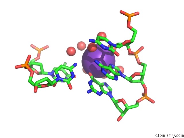
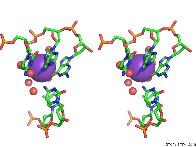
Potassium binding site 1 out of 4 in 4cn5
Go back to
Potassium binding site 1 out
of 4 in the Crystal Structure of the Human Retinoid X Receptor Dna- Binding Domain Bound to the Human NR1D1 Response Element
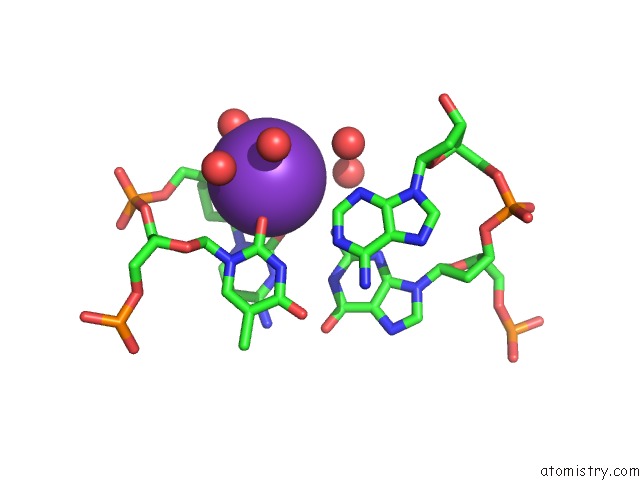
Mono view
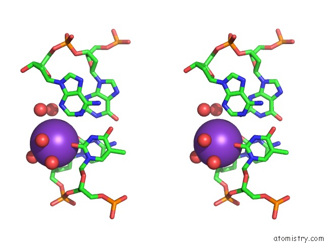
Stereo pair view
Mono view
Stereo pair view
A full contact list of Potassium with other atoms in the K binding
site number 1 of Crystal Structure of the Human Retinoid X Receptor Dna- Binding Domain Bound to the Human NR1D1 Response Element within 5.0Å range:
|
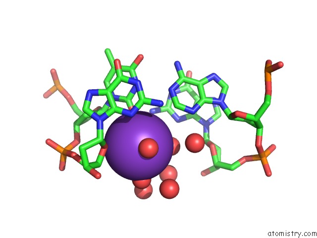
Potassium binding site 2 out of 4 in 4cn5
Go back to
Potassium binding site 2 out
of 4 in the Crystal Structure of the Human Retinoid X Receptor Dna- Binding Domain Bound to the Human NR1D1 Response Element
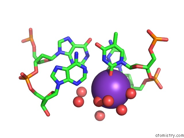
Mono view
Stereo pair view
Mono view
Stereo pair view
A full contact list of Potassium with other atoms in the K binding
site number 2 of Crystal Structure of the Human Retinoid X Receptor Dna- Binding Domain Bound to the Human NR1D1 Response Element within 5.0Å range:
|
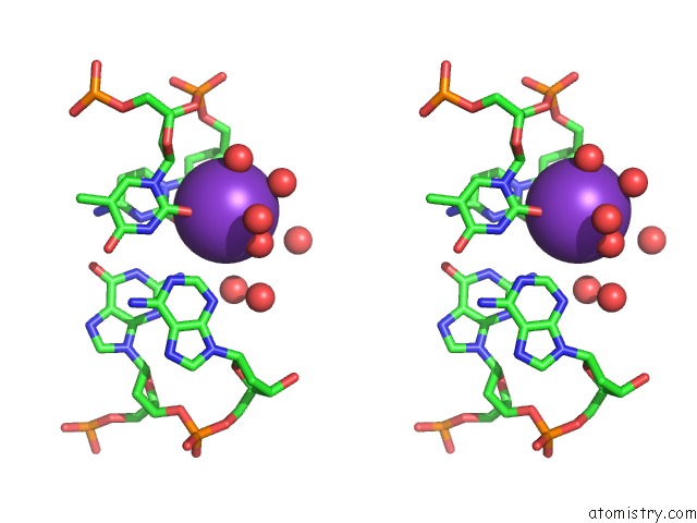
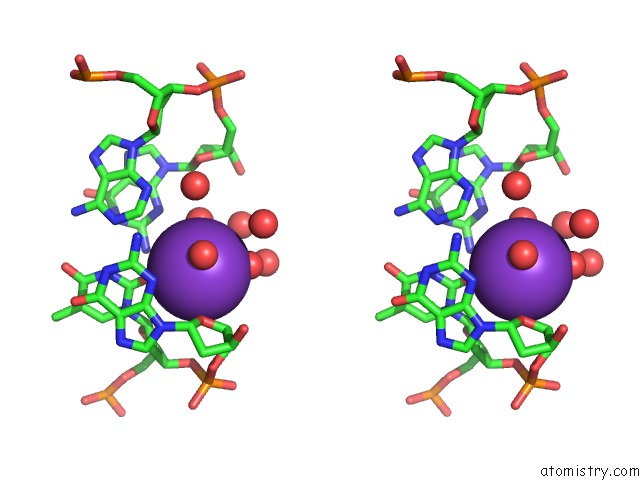
Potassium binding site 3 out of 4 in 4cn5
Go back to
Potassium binding site 3 out
of 4 in the Crystal Structure of the Human Retinoid X Receptor Dna- Binding Domain Bound to the Human NR1D1 Response Element
Mono view
Stereo pair view
Mono view
Stereo pair view
A full contact list of Potassium with other atoms in the K binding
site number 3 of Crystal Structure of the Human Retinoid X Receptor Dna- Binding Domain Bound to the Human NR1D1 Response Element within 5.0Å range:
|
Potassium binding site 4 out of 4 in 4cn5
Go back to
Potassium binding site 4 out
of 4 in the Crystal Structure of the Human Retinoid X Receptor Dna- Binding Domain Bound to the Human NR1D1 Response Element
Mono view
Stereo pair view
Mono view
Stereo pair view
A full contact list of Potassium with other atoms in the K binding
site number 4 of Crystal Structure of the Human Retinoid X Receptor Dna- Binding Domain Bound to the Human NR1D1 Response Element within 5.0Å range:
|
Reference:
J.Osz,
A.G.Mcewen,
P.Poussin-Courmontagne,
E.Moutier,
C.Birck,
I.Davidson,
D.Moras,
N.Rochel.
Structural Basis of Natural Promoter Recognition By the Retinoid X Nuclear Receptor. Sci.Rep. V. 5 8216 2015.
ISSN: ISSN 2045-2322
PubMed: 25645674
DOI: 10.1038/SREP08216
Page generated: Mon Aug 12 10:28:56 2024
ISSN: ISSN 2045-2322
PubMed: 25645674
DOI: 10.1038/SREP08216
Last articles
Zn in 9MJ5Zn in 9HNW
Zn in 9G0L
Zn in 9FNE
Zn in 9DZN
Zn in 9E0I
Zn in 9D32
Zn in 9DAK
Zn in 8ZXC
Zn in 8ZUF