Potassium »
PDB 3zq6-4bg9 »
4av6 »
Potassium in PDB 4av6: Crystal Structure of Thermotoga Maritima Sodium Pumping Membrane Integral Pyrophosphatase at 4 A in Complex with Phosphate and Magnesium
Enzymatic activity of Crystal Structure of Thermotoga Maritima Sodium Pumping Membrane Integral Pyrophosphatase at 4 A in Complex with Phosphate and Magnesium
All present enzymatic activity of Crystal Structure of Thermotoga Maritima Sodium Pumping Membrane Integral Pyrophosphatase at 4 A in Complex with Phosphate and Magnesium:
3.6.1.1;
3.6.1.1;
Protein crystallography data
The structure of Crystal Structure of Thermotoga Maritima Sodium Pumping Membrane Integral Pyrophosphatase at 4 A in Complex with Phosphate and Magnesium, PDB code: 4av6
was solved by
T.Kajander,
J.Kellosalo,
K.Kogan,
K.Pokharel,
A.Goldman,
with X-Ray Crystallography technique. A brief refinement statistics is given in the table below:
| Resolution Low / High (Å) | 29.832 / 4.00 |
| Space group | P 1 21 1 |
| Cell size a, b, c (Å), α, β, γ (°) | 85.800, 112.266, 109.714, 90.00, 103.00, 90.00 |
| R / Rfree (%) | 29.52 / 36.5 |
Other elements in 4av6:
The structure of Crystal Structure of Thermotoga Maritima Sodium Pumping Membrane Integral Pyrophosphatase at 4 A in Complex with Phosphate and Magnesium also contains other interesting chemical elements:
| Magnesium | (Mg) | 8 atoms |
Potassium Binding Sites:
The binding sites of Potassium atom in the Crystal Structure of Thermotoga Maritima Sodium Pumping Membrane Integral Pyrophosphatase at 4 A in Complex with Phosphate and Magnesium
(pdb code 4av6). This binding sites where shown within
5.0 Angstroms radius around Potassium atom.
In total 2 binding sites of Potassium where determined in the Crystal Structure of Thermotoga Maritima Sodium Pumping Membrane Integral Pyrophosphatase at 4 A in Complex with Phosphate and Magnesium, PDB code: 4av6:
Jump to Potassium binding site number: 1; 2;
In total 2 binding sites of Potassium where determined in the Crystal Structure of Thermotoga Maritima Sodium Pumping Membrane Integral Pyrophosphatase at 4 A in Complex with Phosphate and Magnesium, PDB code: 4av6:
Jump to Potassium binding site number: 1; 2;
Potassium binding site 1 out of 2 in 4av6
Go back to
Potassium binding site 1 out
of 2 in the Crystal Structure of Thermotoga Maritima Sodium Pumping Membrane Integral Pyrophosphatase at 4 A in Complex with Phosphate and Magnesium
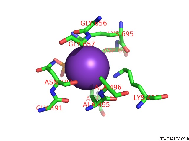
Mono view
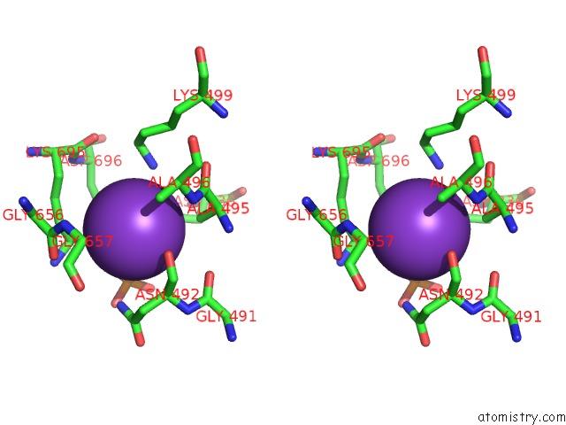
Stereo pair view
Mono view
Stereo pair view
A full contact list of Potassium with other atoms in the K binding
site number 1 of Crystal Structure of Thermotoga Maritima Sodium Pumping Membrane Integral Pyrophosphatase at 4 A in Complex with Phosphate and Magnesium within 5.0Å range:
|
Potassium binding site 2 out of 2 in 4av6
Go back to
Potassium binding site 2 out
of 2 in the Crystal Structure of Thermotoga Maritima Sodium Pumping Membrane Integral Pyrophosphatase at 4 A in Complex with Phosphate and Magnesium
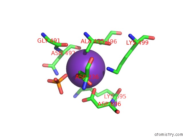
Mono view
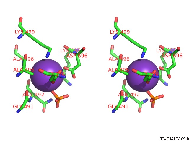
Stereo pair view
Mono view
Stereo pair view
A full contact list of Potassium with other atoms in the K binding
site number 2 of Crystal Structure of Thermotoga Maritima Sodium Pumping Membrane Integral Pyrophosphatase at 4 A in Complex with Phosphate and Magnesium within 5.0Å range:
|
Reference:
J.Kellosalo,
T.Kajander,
K.Kogan,
K.Pokharel,
A.Goldman.
The Structure and Catalytic Cycle of A Sodium-Pumping Pyrophosphatase. Science V. 337 473 2012.
ISSN: ISSN 0036-8075
PubMed: 22837527
DOI: 10.1126/SCIENCE.1222505
Page generated: Mon Aug 12 10:04:19 2024
ISSN: ISSN 0036-8075
PubMed: 22837527
DOI: 10.1126/SCIENCE.1222505
Last articles
Zn in 9MJ5Zn in 9HNW
Zn in 9G0L
Zn in 9FNE
Zn in 9DZN
Zn in 9E0I
Zn in 9D32
Zn in 9DAK
Zn in 8ZXC
Zn in 8ZUF