Potassium »
PDB 3vuw-3zns »
3wnw »
Potassium in PDB 3wnw: Structure of Mouse H-Chain Modified Ferritin
Enzymatic activity of Structure of Mouse H-Chain Modified Ferritin
All present enzymatic activity of Structure of Mouse H-Chain Modified Ferritin:
1.16.3.1;
1.16.3.1;
Protein crystallography data
The structure of Structure of Mouse H-Chain Modified Ferritin, PDB code: 3wnw
was solved by
R.Zarivach,
L.Lewin,
with X-Ray Crystallography technique. A brief refinement statistics is given in the table below:
| Resolution Low / High (Å) | 49.86 / 2.24 |
| Space group | P 42 21 2 |
| Cell size a, b, c (Å), α, β, γ (°) | 218.145, 218.145, 147.690, 90.00, 90.00, 90.00 |
| R / Rfree (%) | 22 / 26.5 |
Other elements in 3wnw:
The structure of Structure of Mouse H-Chain Modified Ferritin also contains other interesting chemical elements:
| Magnesium | (Mg) | 20 atoms |
| Iron | (Fe) | 4 atoms |
Potassium Binding Sites:
The binding sites of Potassium atom in the Structure of Mouse H-Chain Modified Ferritin
(pdb code 3wnw). This binding sites where shown within
5.0 Angstroms radius around Potassium atom.
In total 2 binding sites of Potassium where determined in the Structure of Mouse H-Chain Modified Ferritin, PDB code: 3wnw:
Jump to Potassium binding site number: 1; 2;
In total 2 binding sites of Potassium where determined in the Structure of Mouse H-Chain Modified Ferritin, PDB code: 3wnw:
Jump to Potassium binding site number: 1; 2;
Potassium binding site 1 out of 2 in 3wnw
Go back to
Potassium binding site 1 out
of 2 in the Structure of Mouse H-Chain Modified Ferritin
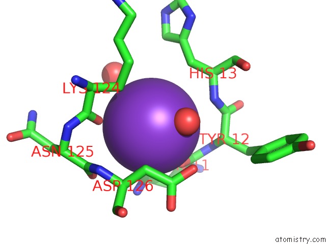
Mono view
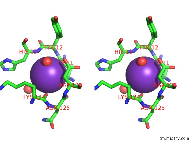
Stereo pair view
Mono view
Stereo pair view
A full contact list of Potassium with other atoms in the K binding
site number 1 of Structure of Mouse H-Chain Modified Ferritin within 5.0Å range:
|
Potassium binding site 2 out of 2 in 3wnw
Go back to
Potassium binding site 2 out
of 2 in the Structure of Mouse H-Chain Modified Ferritin
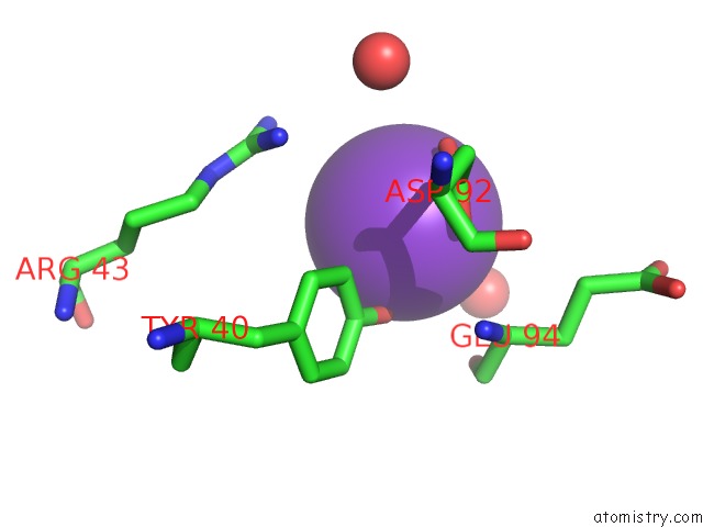
Mono view

Stereo pair view
Mono view
Stereo pair view
A full contact list of Potassium with other atoms in the K binding
site number 2 of Structure of Mouse H-Chain Modified Ferritin within 5.0Å range:
|
Reference:
R.Zarivach,
L.Lewin.
Studies of Ferritin-M6A To Be Published.
Page generated: Sat Aug 9 06:08:26 2025
Last articles
Mg in 4G0VMg in 4G0U
Mg in 4G0R
Mg in 4FXF
Mg in 4FYY
Mg in 4G0N
Mg in 4FZL
Mg in 4FYP
Mg in 4FYX
Mg in 4FVA