Potassium »
PDB 3ukm-3vrs »
3vgm »
Potassium in PDB 3vgm: Crystal Structure of A Rok Family Glucokinase From Streptomyces Griseus in Complex with Glucose
Enzymatic activity of Crystal Structure of A Rok Family Glucokinase From Streptomyces Griseus in Complex with Glucose
All present enzymatic activity of Crystal Structure of A Rok Family Glucokinase From Streptomyces Griseus in Complex with Glucose:
2.7.1.2;
2.7.1.2;
Protein crystallography data
The structure of Crystal Structure of A Rok Family Glucokinase From Streptomyces Griseus in Complex with Glucose, PDB code: 3vgm
was solved by
K.Miyazono,
N.Tabei,
S.Morita,
Y.Ohnishi,
S.Horinouchi,
M.Tanokura,
with X-Ray Crystallography technique. A brief refinement statistics is given in the table below:
| Resolution Low / High (Å) | 20.00 / 1.84 |
| Space group | P 64 2 2 |
| Cell size a, b, c (Å), α, β, γ (°) | 108.190, 108.190, 141.180, 90.00, 90.00, 120.00 |
| R / Rfree (%) | 16.7 / 18.5 |
Other elements in 3vgm:
The structure of Crystal Structure of A Rok Family Glucokinase From Streptomyces Griseus in Complex with Glucose also contains other interesting chemical elements:
| Zinc | (Zn) | 1 atom |
Potassium Binding Sites:
The binding sites of Potassium atom in the Crystal Structure of A Rok Family Glucokinase From Streptomyces Griseus in Complex with Glucose
(pdb code 3vgm). This binding sites where shown within
5.0 Angstroms radius around Potassium atom.
In total only one binding site of Potassium was determined in the Crystal Structure of A Rok Family Glucokinase From Streptomyces Griseus in Complex with Glucose, PDB code: 3vgm:
In total only one binding site of Potassium was determined in the Crystal Structure of A Rok Family Glucokinase From Streptomyces Griseus in Complex with Glucose, PDB code: 3vgm:
Potassium binding site 1 out of 1 in 3vgm
Go back to
Potassium binding site 1 out
of 1 in the Crystal Structure of A Rok Family Glucokinase From Streptomyces Griseus in Complex with Glucose
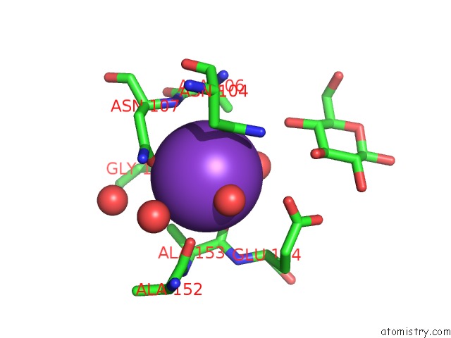
Mono view
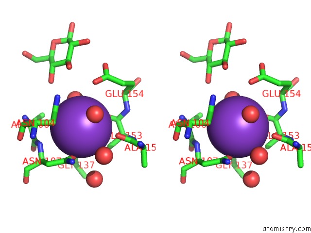
Stereo pair view
Mono view
Stereo pair view
A full contact list of Potassium with other atoms in the K binding
site number 1 of Crystal Structure of A Rok Family Glucokinase From Streptomyces Griseus in Complex with Glucose within 5.0Å range:
|
Reference:
K.Miyazono,
N.Tabei,
S.Morita,
Y.Ohnishi,
S.Horinouchi,
M.Tanokura.
Substrate Recognition Mechanism and Substrate-Dependent Conformational Changes of An Rok Family Glucokinase From Streptomyces Griseus J.Bacteriol. V. 194 607 2012.
ISSN: ISSN 0021-9193
PubMed: 22101842
DOI: 10.1128/JB.06173-11
Page generated: Mon Aug 12 09:48:43 2024
ISSN: ISSN 0021-9193
PubMed: 22101842
DOI: 10.1128/JB.06173-11
Last articles
Zn in 9J0NZn in 9J0O
Zn in 9J0P
Zn in 9FJX
Zn in 9EKB
Zn in 9C0F
Zn in 9CAH
Zn in 9CH0
Zn in 9CH3
Zn in 9CH1