Potassium »
PDB 3ukm-3vrs »
3usz »
Potassium in PDB 3usz: Crystal Structure of Truncated Exo-1,3/1,4-Beta-Glucanase (Exop) From Pseudoalteromonas Sp. BB1
Protein crystallography data
The structure of Crystal Structure of Truncated Exo-1,3/1,4-Beta-Glucanase (Exop) From Pseudoalteromonas Sp. BB1, PDB code: 3usz
was solved by
Y.Nakatani,
S.M.Cutfield,
J.F.Cutfield,
with X-Ray Crystallography technique. A brief refinement statistics is given in the table below:
| Resolution Low / High (Å) | 33.42 / 2.10 |
| Space group | C 2 2 21 |
| Cell size a, b, c (Å), α, β, γ (°) | 68.054, 179.414, 177.177, 90.00, 90.00, 90.00 |
| R / Rfree (%) | 16.4 / 19.4 |
Other elements in 3usz:
The structure of Crystal Structure of Truncated Exo-1,3/1,4-Beta-Glucanase (Exop) From Pseudoalteromonas Sp. BB1 also contains other interesting chemical elements:
| Calcium | (Ca) | 1 atom |
| Sodium | (Na) | 1 atom |
Potassium Binding Sites:
The binding sites of Potassium atom in the Crystal Structure of Truncated Exo-1,3/1,4-Beta-Glucanase (Exop) From Pseudoalteromonas Sp. BB1
(pdb code 3usz). This binding sites where shown within
5.0 Angstroms radius around Potassium atom.
In total only one binding site of Potassium was determined in the Crystal Structure of Truncated Exo-1,3/1,4-Beta-Glucanase (Exop) From Pseudoalteromonas Sp. BB1, PDB code: 3usz:
In total only one binding site of Potassium was determined in the Crystal Structure of Truncated Exo-1,3/1,4-Beta-Glucanase (Exop) From Pseudoalteromonas Sp. BB1, PDB code: 3usz:
Potassium binding site 1 out of 1 in 3usz
Go back to
Potassium binding site 1 out
of 1 in the Crystal Structure of Truncated Exo-1,3/1,4-Beta-Glucanase (Exop) From Pseudoalteromonas Sp. BB1
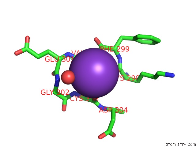
Mono view
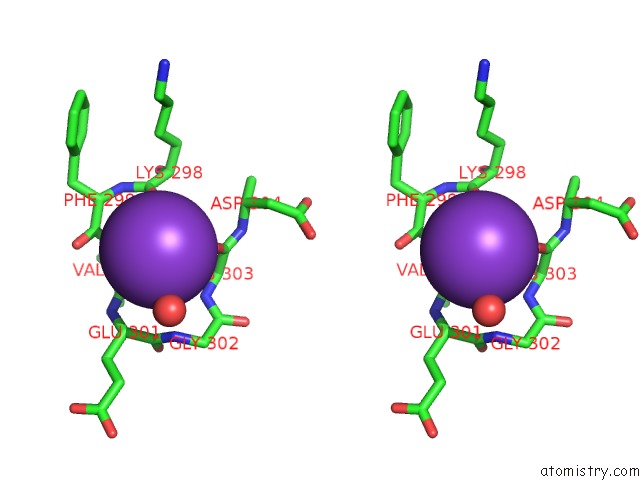
Stereo pair view
Mono view
Stereo pair view
A full contact list of Potassium with other atoms in the K binding
site number 1 of Crystal Structure of Truncated Exo-1,3/1,4-Beta-Glucanase (Exop) From Pseudoalteromonas Sp. BB1 within 5.0Å range:
|
Reference:
Y.Nakatani,
S.M.Cutfield,
N.P.Cowieson,
J.F.Cutfield.
Structure and Activity of Exo-1,3/1,4-Beta-Glucanase From Marine Bacterium Pseudoalteromonas Sp. BB1 Showing A Novel C-Terminal Domain Febs J. 2011.
ISSN: ISSN 1742-464X
PubMed: 22129429
DOI: 10.1111/J.1742-4658.2011.08439.X
Page generated: Mon Aug 12 09:43:34 2024
ISSN: ISSN 1742-464X
PubMed: 22129429
DOI: 10.1111/J.1742-4658.2011.08439.X
Last articles
Zn in 9J0NZn in 9J0O
Zn in 9J0P
Zn in 9FJX
Zn in 9EKB
Zn in 9C0F
Zn in 9CAH
Zn in 9CH0
Zn in 9CH3
Zn in 9CH1