Potassium »
PDB 3ukm-3vrs »
3umo »
Potassium in PDB 3umo: Crystal Structure of the Phosphofructokinase-2 From Escherichia Coli in Complex with Potassium
Enzymatic activity of Crystal Structure of the Phosphofructokinase-2 From Escherichia Coli in Complex with Potassium
All present enzymatic activity of Crystal Structure of the Phosphofructokinase-2 From Escherichia Coli in Complex with Potassium:
2.7.1.11;
2.7.1.11;
Protein crystallography data
The structure of Crystal Structure of the Phosphofructokinase-2 From Escherichia Coli in Complex with Potassium, PDB code: 3umo
was solved by
H.M.Pereira,
A.Caniuguir,
M.Baez,
R.Cabrera,
R.C.Garratt,
J.Babul,
with X-Ray Crystallography technique. A brief refinement statistics is given in the table below:
| Resolution Low / High (Å) | 48.97 / 1.70 |
| Space group | P 2 2 21 |
| Cell size a, b, c (Å), α, β, γ (°) | 43.812, 88.770, 176.120, 90.00, 90.00, 90.00 |
| R / Rfree (%) | 18.1 / 20.8 |
Other elements in 3umo:
The structure of Crystal Structure of the Phosphofructokinase-2 From Escherichia Coli in Complex with Potassium also contains other interesting chemical elements:
| Magnesium | (Mg) | 4 atoms |
Potassium Binding Sites:
The binding sites of Potassium atom in the Crystal Structure of the Phosphofructokinase-2 From Escherichia Coli in Complex with Potassium
(pdb code 3umo). This binding sites where shown within
5.0 Angstroms radius around Potassium atom.
In total 2 binding sites of Potassium where determined in the Crystal Structure of the Phosphofructokinase-2 From Escherichia Coli in Complex with Potassium, PDB code: 3umo:
Jump to Potassium binding site number: 1; 2;
In total 2 binding sites of Potassium where determined in the Crystal Structure of the Phosphofructokinase-2 From Escherichia Coli in Complex with Potassium, PDB code: 3umo:
Jump to Potassium binding site number: 1; 2;
Potassium binding site 1 out of 2 in 3umo
Go back to
Potassium binding site 1 out
of 2 in the Crystal Structure of the Phosphofructokinase-2 From Escherichia Coli in Complex with Potassium
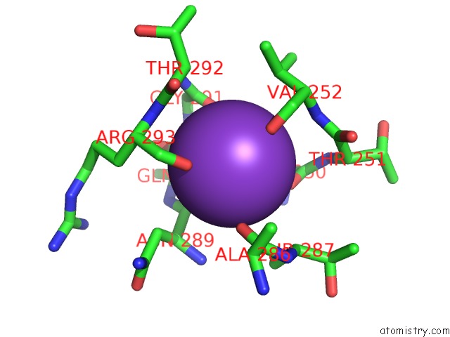
Mono view
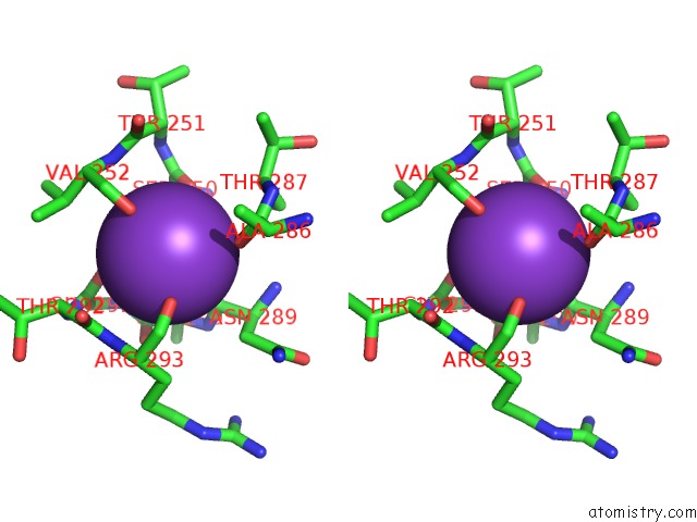
Stereo pair view
Mono view
Stereo pair view
A full contact list of Potassium with other atoms in the K binding
site number 1 of Crystal Structure of the Phosphofructokinase-2 From Escherichia Coli in Complex with Potassium within 5.0Å range:
|
Potassium binding site 2 out of 2 in 3umo
Go back to
Potassium binding site 2 out
of 2 in the Crystal Structure of the Phosphofructokinase-2 From Escherichia Coli in Complex with Potassium
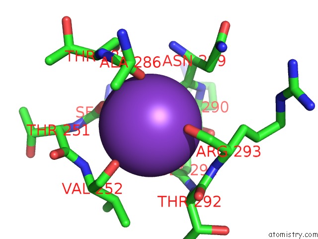
Mono view
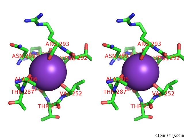
Stereo pair view
Mono view
Stereo pair view
A full contact list of Potassium with other atoms in the K binding
site number 2 of Crystal Structure of the Phosphofructokinase-2 From Escherichia Coli in Complex with Potassium within 5.0Å range:
|
Reference:
M.Baez,
R.Cabrera,
H.M.Pereira,
A.Blanco,
P.Villalobos,
C.A.Ramirez-Sarmiento,
A.Caniuguir,
V.Guixe,
R.C.Garratt,
J.Babul.
A Ribokinase Family Conserved Monovalent Cation Binding Site Enhances the Mgatp-Induced Inhibition in E. Coli Phosphofructokinase-2 Biophys.J. V. 105 185 2013.
ISSN: ISSN 0006-3495
PubMed: 23823238
DOI: 10.1016/J.BPJ.2013.05.028
Page generated: Sat Aug 9 06:00:48 2025
ISSN: ISSN 0006-3495
PubMed: 23823238
DOI: 10.1016/J.BPJ.2013.05.028
Last articles
K in 7OV7K in 7OTJ
K in 7OTB
K in 7OPH
K in 7OUP
K in 7OUE
K in 7OQT
K in 7OQ1
K in 7OT4
K in 7OOU