Potassium »
PDB 3srd-3ugx »
3u81 »
Potassium in PDB 3u81: Crystal Structure of A Sah-Bound Semi-Holo Form of Rat Catechol-O- Methyltransferase
Enzymatic activity of Crystal Structure of A Sah-Bound Semi-Holo Form of Rat Catechol-O- Methyltransferase
All present enzymatic activity of Crystal Structure of A Sah-Bound Semi-Holo Form of Rat Catechol-O- Methyltransferase:
2.1.1.6;
2.1.1.6;
Protein crystallography data
The structure of Crystal Structure of A Sah-Bound Semi-Holo Form of Rat Catechol-O- Methyltransferase, PDB code: 3u81
was solved by
A.Ehler,
D.Schlatter,
M.Stihle,
J.Benz,
M.G.Rudolph,
with X-Ray Crystallography technique. A brief refinement statistics is given in the table below:
| Resolution Low / High (Å) | 39.81 / 1.13 |
| Space group | P 21 21 21 |
| Cell size a, b, c (Å), α, β, γ (°) | 33.384, 61.333, 104.686, 90.00, 90.00, 90.00 |
| R / Rfree (%) | 13.4 / 16 |
Potassium Binding Sites:
The binding sites of Potassium atom in the Crystal Structure of A Sah-Bound Semi-Holo Form of Rat Catechol-O- Methyltransferase
(pdb code 3u81). This binding sites where shown within
5.0 Angstroms radius around Potassium atom.
In total only one binding site of Potassium was determined in the Crystal Structure of A Sah-Bound Semi-Holo Form of Rat Catechol-O- Methyltransferase, PDB code: 3u81:
In total only one binding site of Potassium was determined in the Crystal Structure of A Sah-Bound Semi-Holo Form of Rat Catechol-O- Methyltransferase, PDB code: 3u81:
Potassium binding site 1 out of 1 in 3u81
Go back to
Potassium binding site 1 out
of 1 in the Crystal Structure of A Sah-Bound Semi-Holo Form of Rat Catechol-O- Methyltransferase
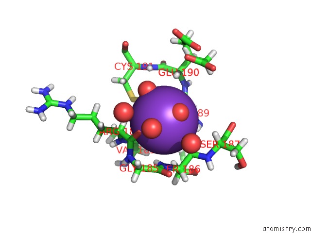
Mono view
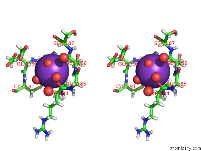
Stereo pair view
Mono view
Stereo pair view
A full contact list of Potassium with other atoms in the K binding
site number 1 of Crystal Structure of A Sah-Bound Semi-Holo Form of Rat Catechol-O- Methyltransferase within 5.0Å range:
|
Reference:
M.Ellermann,
C.Lerner,
G.Burgy,
A.Ehler,
C.Bissantz,
R.Jakob-Roetne,
R.Paulini,
O.Allemann,
H.Tissot,
D.Grunstein,
M.Stihle,
F.Diederich,
M.G.Rudolph.
Catechol-O-Methyltransferase in Complex with Substituted 3'-Deoxyribose Bisubstrate Inhibitors. Acta Crystallogr.,Sect.D V. 68 253 2012.
ISSN: ISSN 0907-4449
PubMed: 22349227
DOI: 10.1107/S0907444912001138
Page generated: Mon Aug 12 09:41:40 2024
ISSN: ISSN 0907-4449
PubMed: 22349227
DOI: 10.1107/S0907444912001138
Last articles
Zn in 9JYWZn in 9IR4
Zn in 9IR3
Zn in 9GMX
Zn in 9GMW
Zn in 9JEJ
Zn in 9ERF
Zn in 9ERE
Zn in 9EGV
Zn in 9EGW