Potassium »
PDB 3srd-3ugx »
3syc »
Potassium in PDB 3syc: Crystal Structure of the G Protein-Gated Inward Rectifier K+ Channel GIRK2 (KIR3.2) D228N Mutant
Protein crystallography data
The structure of Crystal Structure of the G Protein-Gated Inward Rectifier K+ Channel GIRK2 (KIR3.2) D228N Mutant, PDB code: 3syc
was solved by
M.R.Whorton,
R.Mackinnon,
with X-Ray Crystallography technique. A brief refinement statistics is given in the table below:
| Resolution Low / High (Å) | 42.04 / 3.41 |
| Space group | P 4 21 2 |
| Cell size a, b, c (Å), α, β, γ (°) | 86.488, 86.488, 179.627, 90.00, 90.00, 90.00 |
| R / Rfree (%) | 25.1 / 27.1 |
Potassium Binding Sites:
The binding sites of Potassium atom in the Crystal Structure of the G Protein-Gated Inward Rectifier K+ Channel GIRK2 (KIR3.2) D228N Mutant
(pdb code 3syc). This binding sites where shown within
5.0 Angstroms radius around Potassium atom.
In total 6 binding sites of Potassium where determined in the Crystal Structure of the G Protein-Gated Inward Rectifier K+ Channel GIRK2 (KIR3.2) D228N Mutant, PDB code: 3syc:
Jump to Potassium binding site number: 1; 2; 3; 4; 5; 6;
In total 6 binding sites of Potassium where determined in the Crystal Structure of the G Protein-Gated Inward Rectifier K+ Channel GIRK2 (KIR3.2) D228N Mutant, PDB code: 3syc:
Jump to Potassium binding site number: 1; 2; 3; 4; 5; 6;
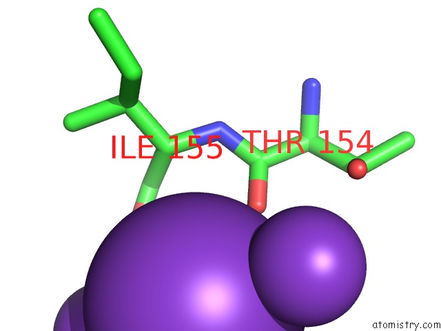
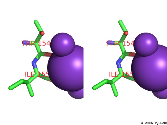
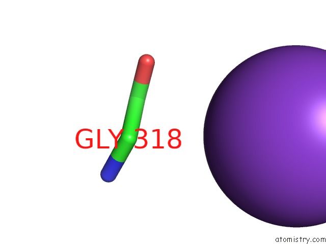
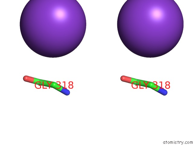
Potassium binding site 1 out of 6 in 3syc
Go back to
Potassium binding site 1 out
of 6 in the Crystal Structure of the G Protein-Gated Inward Rectifier K+ Channel GIRK2 (KIR3.2) D228N Mutant
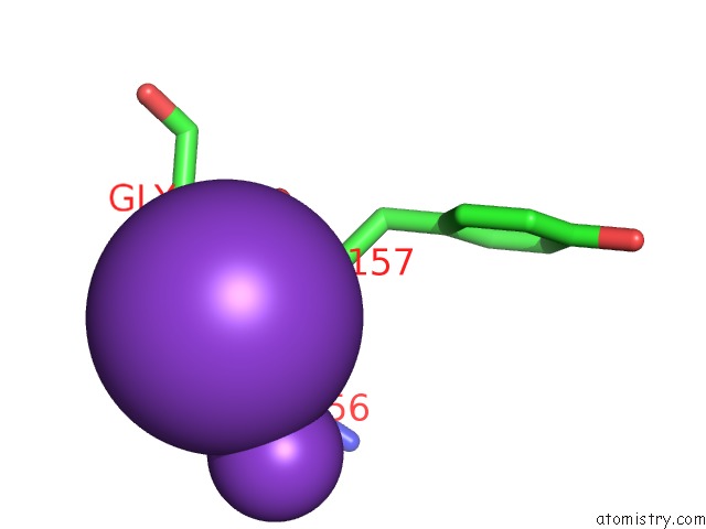
Mono view
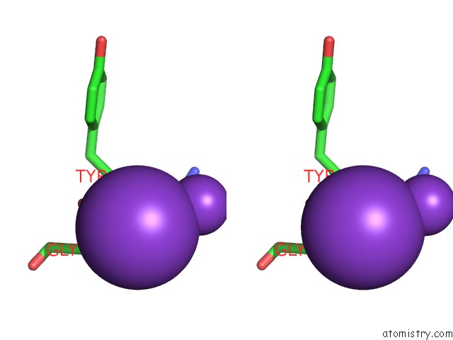
Stereo pair view
Mono view
Stereo pair view
A full contact list of Potassium with other atoms in the K binding
site number 1 of Crystal Structure of the G Protein-Gated Inward Rectifier K+ Channel GIRK2 (KIR3.2) D228N Mutant within 5.0Å range:
|
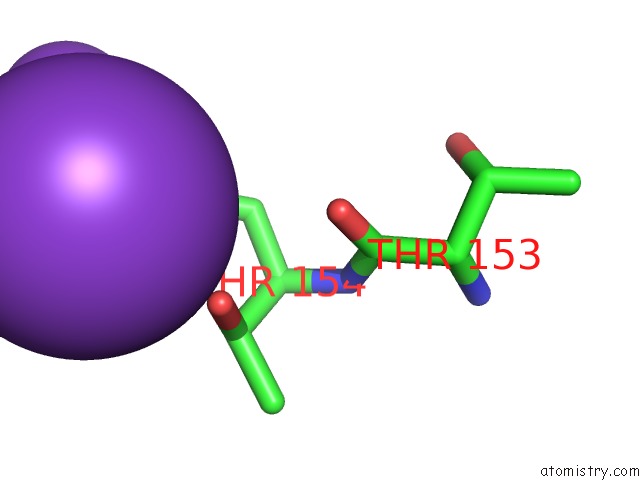
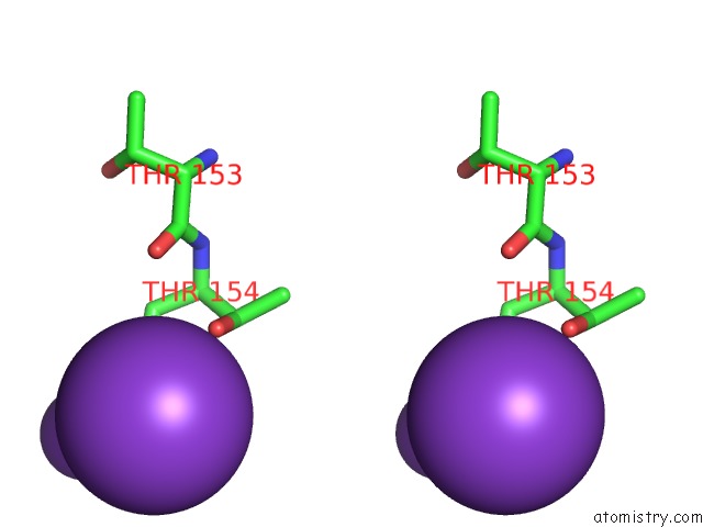
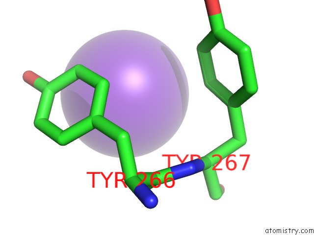
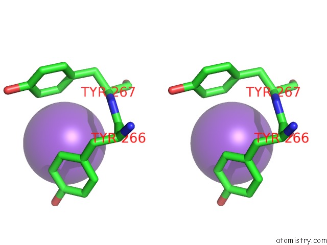
Potassium binding site 2 out of 6 in 3syc
Go back to
Potassium binding site 2 out
of 6 in the Crystal Structure of the G Protein-Gated Inward Rectifier K+ Channel GIRK2 (KIR3.2) D228N Mutant
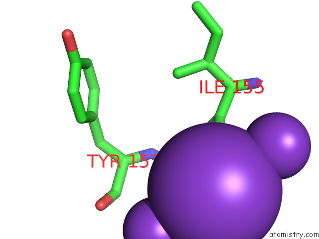
Mono view
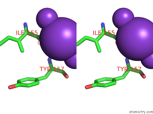
Stereo pair view
Mono view
Stereo pair view
A full contact list of Potassium with other atoms in the K binding
site number 2 of Crystal Structure of the G Protein-Gated Inward Rectifier K+ Channel GIRK2 (KIR3.2) D228N Mutant within 5.0Å range:
|
Potassium binding site 3 out of 6 in 3syc
Go back to
Potassium binding site 3 out
of 6 in the Crystal Structure of the G Protein-Gated Inward Rectifier K+ Channel GIRK2 (KIR3.2) D228N Mutant
Mono view
Stereo pair view
Mono view
Stereo pair view
A full contact list of Potassium with other atoms in the K binding
site number 3 of Crystal Structure of the G Protein-Gated Inward Rectifier K+ Channel GIRK2 (KIR3.2) D228N Mutant within 5.0Å range:
|
Potassium binding site 4 out of 6 in 3syc
Go back to
Potassium binding site 4 out
of 6 in the Crystal Structure of the G Protein-Gated Inward Rectifier K+ Channel GIRK2 (KIR3.2) D228N Mutant
Mono view
Stereo pair view
Mono view
Stereo pair view
A full contact list of Potassium with other atoms in the K binding
site number 4 of Crystal Structure of the G Protein-Gated Inward Rectifier K+ Channel GIRK2 (KIR3.2) D228N Mutant within 5.0Å range:
|
Potassium binding site 5 out of 6 in 3syc
Go back to
Potassium binding site 5 out
of 6 in the Crystal Structure of the G Protein-Gated Inward Rectifier K+ Channel GIRK2 (KIR3.2) D228N Mutant
Mono view
Stereo pair view
Mono view
Stereo pair view
A full contact list of Potassium with other atoms in the K binding
site number 5 of Crystal Structure of the G Protein-Gated Inward Rectifier K+ Channel GIRK2 (KIR3.2) D228N Mutant within 5.0Å range:
|
Potassium binding site 6 out of 6 in 3syc
Go back to
Potassium binding site 6 out
of 6 in the Crystal Structure of the G Protein-Gated Inward Rectifier K+ Channel GIRK2 (KIR3.2) D228N Mutant
Mono view
Stereo pair view
Mono view
Stereo pair view
A full contact list of Potassium with other atoms in the K binding
site number 6 of Crystal Structure of the G Protein-Gated Inward Rectifier K+ Channel GIRK2 (KIR3.2) D228N Mutant within 5.0Å range:
|
Reference:
M.R.Whorton,
R.Mackinnon.
Crystal Structure of the Mammalian GIRK2 K(+) Channel and Gating Regulation By G Proteins, Pip(2), and Sodium. Cell(Cambridge,Mass.) V. 147 199 2011.
ISSN: ISSN 0092-8674
PubMed: 21962516
DOI: 10.1016/J.CELL.2011.07.046
Page generated: Mon Aug 12 09:33:58 2024
ISSN: ISSN 0092-8674
PubMed: 21962516
DOI: 10.1016/J.CELL.2011.07.046
Last articles
Zn in 9JYWZn in 9IR4
Zn in 9IR3
Zn in 9GMX
Zn in 9GMW
Zn in 9JEJ
Zn in 9ERF
Zn in 9ERE
Zn in 9EGV
Zn in 9EGW