Potassium »
PDB 3rs9-3spj »
3s7k »
Potassium in PDB 3s7k: Structure of Thrombin Mutant Y225P in the E Form
Enzymatic activity of Structure of Thrombin Mutant Y225P in the E Form
All present enzymatic activity of Structure of Thrombin Mutant Y225P in the E Form:
3.4.21.5;
3.4.21.5;
Protein crystallography data
The structure of Structure of Thrombin Mutant Y225P in the E Form, PDB code: 3s7k
was solved by
W.Niu,
Z.Chen,
P.Gandhi,
A.Vogt,
N.Pozzi,
L.A.Pele,
F.Zapata,
E.Di Cera,
with X-Ray Crystallography technique. A brief refinement statistics is given in the table below:
| Resolution Low / High (Å) | 35.67 / 1.90 |
| Space group | P 21 21 2 |
| Cell size a, b, c (Å), α, β, γ (°) | 61.937, 86.813, 100.968, 90.00, 90.00, 90.00 |
| R / Rfree (%) | 17.5 / 21.7 |
Potassium Binding Sites:
The binding sites of Potassium atom in the Structure of Thrombin Mutant Y225P in the E Form
(pdb code 3s7k). This binding sites where shown within
5.0 Angstroms radius around Potassium atom.
In total 2 binding sites of Potassium where determined in the Structure of Thrombin Mutant Y225P in the E Form, PDB code: 3s7k:
Jump to Potassium binding site number: 1; 2;
In total 2 binding sites of Potassium where determined in the Structure of Thrombin Mutant Y225P in the E Form, PDB code: 3s7k:
Jump to Potassium binding site number: 1; 2;
Potassium binding site 1 out of 2 in 3s7k
Go back to
Potassium binding site 1 out
of 2 in the Structure of Thrombin Mutant Y225P in the E Form
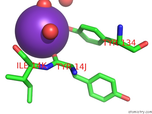
Mono view
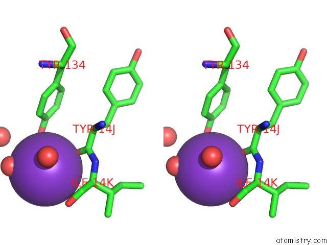
Stereo pair view
Mono view
Stereo pair view
A full contact list of Potassium with other atoms in the K binding
site number 1 of Structure of Thrombin Mutant Y225P in the E Form within 5.0Å range:
|
Potassium binding site 2 out of 2 in 3s7k
Go back to
Potassium binding site 2 out
of 2 in the Structure of Thrombin Mutant Y225P in the E Form
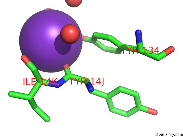
Mono view
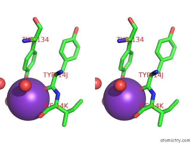
Stereo pair view
Mono view
Stereo pair view
A full contact list of Potassium with other atoms in the K binding
site number 2 of Structure of Thrombin Mutant Y225P in the E Form within 5.0Å range:
|
Reference:
W.Niu,
Z.Chen,
P.S.Gandhi,
A.D.Vogt,
N.Pozzi,
L.A.Pelc,
F.Zapata,
E.Di Cera.
Crystallographic and Kinetic Evidence of Allostery in A Trypsin-Like Protease. Biochemistry V. 50 6301 2011.
ISSN: ISSN 0006-2960
PubMed: 21707111
DOI: 10.1021/BI200878C
Page generated: Mon Aug 12 09:25:47 2024
ISSN: ISSN 0006-2960
PubMed: 21707111
DOI: 10.1021/BI200878C
Last articles
Zn in 9J0NZn in 9J0O
Zn in 9J0P
Zn in 9FJX
Zn in 9EKB
Zn in 9C0F
Zn in 9CAH
Zn in 9CH0
Zn in 9CH3
Zn in 9CH1