Potassium »
PDB 3rs9-3spj »
3s5n »
Potassium in PDB 3s5n: Crystal Structure of Human 4-Hydroxy-2-Oxoglutarate Aldolase
Enzymatic activity of Crystal Structure of Human 4-Hydroxy-2-Oxoglutarate Aldolase
All present enzymatic activity of Crystal Structure of Human 4-Hydroxy-2-Oxoglutarate Aldolase:
4.1.3.16;
4.1.3.16;
Protein crystallography data
The structure of Crystal Structure of Human 4-Hydroxy-2-Oxoglutarate Aldolase, PDB code: 3s5n
was solved by
T.J.Riedel,
W.T.Lowther,
with X-Ray Crystallography technique. A brief refinement statistics is given in the table below:
| Resolution Low / High (Å) | 46.52 / 2.50 |
| Space group | P 64 2 2 |
| Cell size a, b, c (Å), α, β, γ (°) | 142.143, 142.143, 108.070, 90.00, 90.00, 120.00 |
| R / Rfree (%) | 21.2 / 24.6 |
Other elements in 3s5n:
The structure of Crystal Structure of Human 4-Hydroxy-2-Oxoglutarate Aldolase also contains other interesting chemical elements:
| Sodium | (Na) | 1 atom |
Potassium Binding Sites:
The binding sites of Potassium atom in the Crystal Structure of Human 4-Hydroxy-2-Oxoglutarate Aldolase
(pdb code 3s5n). This binding sites where shown within
5.0 Angstroms radius around Potassium atom.
In total only one binding site of Potassium was determined in the Crystal Structure of Human 4-Hydroxy-2-Oxoglutarate Aldolase, PDB code: 3s5n:
In total only one binding site of Potassium was determined in the Crystal Structure of Human 4-Hydroxy-2-Oxoglutarate Aldolase, PDB code: 3s5n:
Potassium binding site 1 out of 1 in 3s5n
Go back to
Potassium binding site 1 out
of 1 in the Crystal Structure of Human 4-Hydroxy-2-Oxoglutarate Aldolase
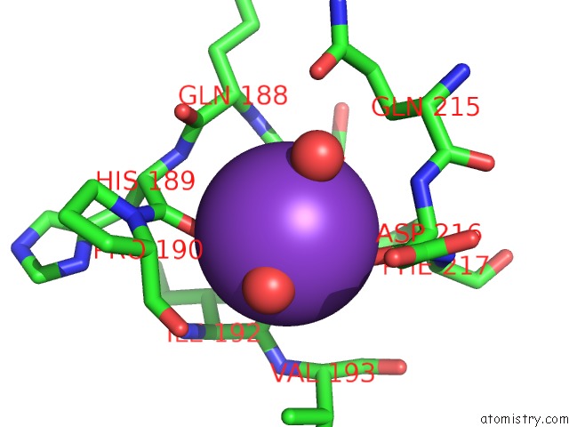
Mono view
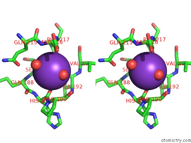
Stereo pair view
Mono view
Stereo pair view
A full contact list of Potassium with other atoms in the K binding
site number 1 of Crystal Structure of Human 4-Hydroxy-2-Oxoglutarate Aldolase within 5.0Å range:
|
Reference:
T.J.Riedel,
L.C.Johnson,
J.Knight,
R.R.Hantgan,
R.P.Holmes,
W.T.Lowther.
Structural and Biochemical Studies of Human 4-Hydroxy-2-Oxoglutarate Aldolase: Implications For Hydroxyproline Metabolism in Primary Hyperoxaluria. Plos One V. 6 26021 2011.
ISSN: ESSN 1932-6203
PubMed: 21998747
DOI: 10.1371/JOURNAL.PONE.0026021
Page generated: Mon Aug 12 09:25:44 2024
ISSN: ESSN 1932-6203
PubMed: 21998747
DOI: 10.1371/JOURNAL.PONE.0026021
Last articles
Zn in 9J0NZn in 9J0O
Zn in 9J0P
Zn in 9FJX
Zn in 9EKB
Zn in 9C0F
Zn in 9CAH
Zn in 9CH0
Zn in 9CH3
Zn in 9CH1