Potassium »
PDB 3q9c-3rs8 »
3qek »
Potassium in PDB 3qek: Crystal Structure of Amino Terminal Domain of the Nmda Receptor Subunit GLUN1
Protein crystallography data
The structure of Crystal Structure of Amino Terminal Domain of the Nmda Receptor Subunit GLUN1, PDB code: 3qek
was solved by
E.Karakas,
N.Simorowski,
H.Furukawa,
with X-Ray Crystallography technique. A brief refinement statistics is given in the table below:
| Resolution Low / High (Å) | 29.95 / 2.00 |
| Space group | P 21 21 21 |
| Cell size a, b, c (Å), α, β, γ (°) | 47.323, 92.813, 209.978, 90.00, 90.00, 90.00 |
| R / Rfree (%) | 19.2 / 22.6 |
Potassium Binding Sites:
The binding sites of Potassium atom in the Crystal Structure of Amino Terminal Domain of the Nmda Receptor Subunit GLUN1
(pdb code 3qek). This binding sites where shown within
5.0 Angstroms radius around Potassium atom.
In total 2 binding sites of Potassium where determined in the Crystal Structure of Amino Terminal Domain of the Nmda Receptor Subunit GLUN1, PDB code: 3qek:
Jump to Potassium binding site number: 1; 2;
In total 2 binding sites of Potassium where determined in the Crystal Structure of Amino Terminal Domain of the Nmda Receptor Subunit GLUN1, PDB code: 3qek:
Jump to Potassium binding site number: 1; 2;
Potassium binding site 1 out of 2 in 3qek
Go back to
Potassium binding site 1 out
of 2 in the Crystal Structure of Amino Terminal Domain of the Nmda Receptor Subunit GLUN1

Mono view
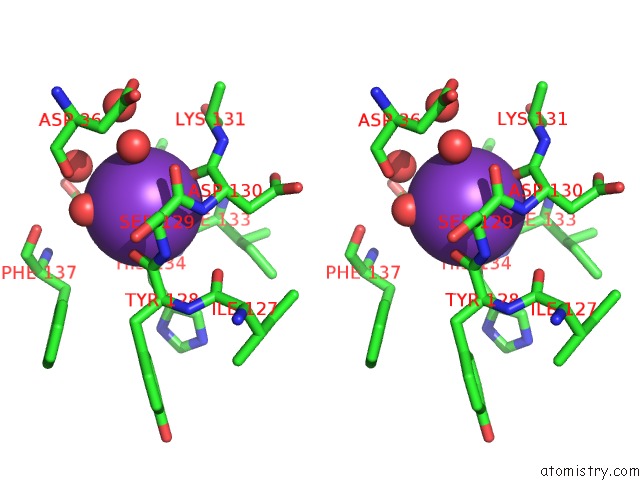
Stereo pair view
Mono view
Stereo pair view
A full contact list of Potassium with other atoms in the K binding
site number 1 of Crystal Structure of Amino Terminal Domain of the Nmda Receptor Subunit GLUN1 within 5.0Å range:
|
Potassium binding site 2 out of 2 in 3qek
Go back to
Potassium binding site 2 out
of 2 in the Crystal Structure of Amino Terminal Domain of the Nmda Receptor Subunit GLUN1
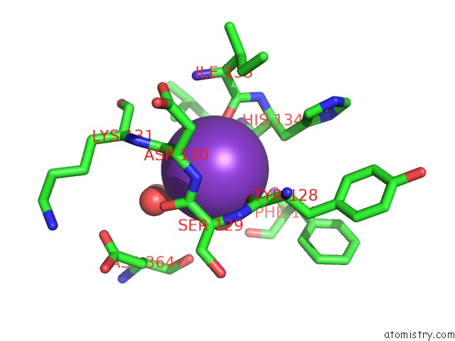
Mono view
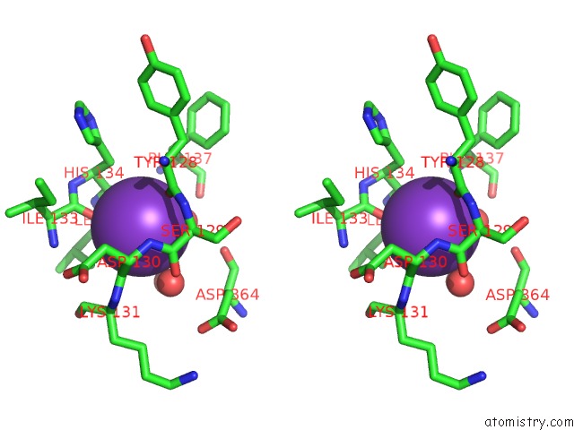
Stereo pair view
Mono view
Stereo pair view
A full contact list of Potassium with other atoms in the K binding
site number 2 of Crystal Structure of Amino Terminal Domain of the Nmda Receptor Subunit GLUN1 within 5.0Å range:
|
Reference:
E.Karakas,
N.Simorowski,
H.Furukawa.
Subunit Arrangement and Phenylethanolamine Binding in GLUN1/GLUN2B Nmda Receptors. Nature V. 475 249 2011.
ISSN: ISSN 0028-0836
PubMed: 21677647
DOI: 10.1038/NATURE10180
Page generated: Mon Aug 12 09:11:22 2024
ISSN: ISSN 0028-0836
PubMed: 21677647
DOI: 10.1038/NATURE10180
Last articles
Zn in 9J0NZn in 9J0O
Zn in 9J0P
Zn in 9FJX
Zn in 9EKB
Zn in 9C0F
Zn in 9CAH
Zn in 9CH0
Zn in 9CH3
Zn in 9CH1