Potassium »
PDB 3m62-3ow2 »
3nal »
Potassium in PDB 3nal: Sr Ca(2+)-Atpase in the HNE2 State Complexed with the Thapsigargin Derivative Dtb
Enzymatic activity of Sr Ca(2+)-Atpase in the HNE2 State Complexed with the Thapsigargin Derivative Dtb
All present enzymatic activity of Sr Ca(2+)-Atpase in the HNE2 State Complexed with the Thapsigargin Derivative Dtb:
3.6.3.8;
3.6.3.8;
Protein crystallography data
The structure of Sr Ca(2+)-Atpase in the HNE2 State Complexed with the Thapsigargin Derivative Dtb, PDB code: 3nal
was solved by
A.M.L.Winther,
Y.Sonntag,
C.Olesen,
J.V.Moller,
P.Nissen,
with X-Ray Crystallography technique. A brief refinement statistics is given in the table below:
| Resolution Low / High (Å) | 50.00 / 2.65 |
| Space group | P 41 21 2 |
| Cell size a, b, c (Å), α, β, γ (°) | 71.330, 71.330, 591.020, 90.00, 90.00, 90.00 |
| R / Rfree (%) | 25.5 / 28.3 |
Other elements in 3nal:
The structure of Sr Ca(2+)-Atpase in the HNE2 State Complexed with the Thapsigargin Derivative Dtb also contains other interesting chemical elements:
| Magnesium | (Mg) | 1 atom |
Potassium Binding Sites:
The binding sites of Potassium atom in the Sr Ca(2+)-Atpase in the HNE2 State Complexed with the Thapsigargin Derivative Dtb
(pdb code 3nal). This binding sites where shown within
5.0 Angstroms radius around Potassium atom.
In total only one binding site of Potassium was determined in the Sr Ca(2+)-Atpase in the HNE2 State Complexed with the Thapsigargin Derivative Dtb, PDB code: 3nal:
In total only one binding site of Potassium was determined in the Sr Ca(2+)-Atpase in the HNE2 State Complexed with the Thapsigargin Derivative Dtb, PDB code: 3nal:
Potassium binding site 1 out of 1 in 3nal
Go back to
Potassium binding site 1 out
of 1 in the Sr Ca(2+)-Atpase in the HNE2 State Complexed with the Thapsigargin Derivative Dtb
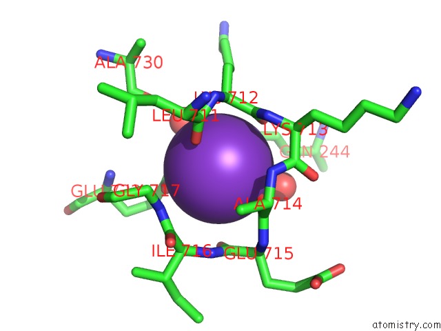
Mono view
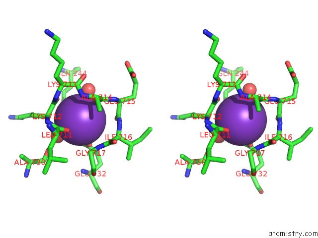
Stereo pair view
Mono view
Stereo pair view
A full contact list of Potassium with other atoms in the K binding
site number 1 of Sr Ca(2+)-Atpase in the HNE2 State Complexed with the Thapsigargin Derivative Dtb within 5.0Å range:
|
Reference:
A.M.L.Winther,
H.Liu,
Y.Sonntag,
C.Olesen,
M.Le Maire,
H.Soehoel,
C.E.Olsen,
S.B.Christensen,
P.Nissen,
J.V.Moller.
Critical Roles of Hydrophobicity and Orientation of Side Chains For Inactivation of Sarcoplasmic Reticulum CA2+-Atpase with Thapsigargin and Thapsigargin Analogs J.Biol.Chem. V. 285 28883 2010.
ISSN: ISSN 0021-9258
PubMed: 20551329
DOI: 10.1074/JBC.M110.136242
Page generated: Mon Aug 12 08:50:02 2024
ISSN: ISSN 0021-9258
PubMed: 20551329
DOI: 10.1074/JBC.M110.136242
Last articles
Zn in 9JYWZn in 9IR4
Zn in 9IR3
Zn in 9GMX
Zn in 9GMW
Zn in 9JEJ
Zn in 9ERF
Zn in 9ERE
Zn in 9EGV
Zn in 9EGW