Potassium »
PDB 3m62-3ow2 »
3n25 »
Potassium in PDB 3n25: The Structure of Muscle Pyruvate Kinase in Complex with Proline, Pyruvate, and MN2+
Enzymatic activity of The Structure of Muscle Pyruvate Kinase in Complex with Proline, Pyruvate, and MN2+
All present enzymatic activity of The Structure of Muscle Pyruvate Kinase in Complex with Proline, Pyruvate, and MN2+:
2.7.1.40;
2.7.1.40;
Protein crystallography data
The structure of The Structure of Muscle Pyruvate Kinase in Complex with Proline, Pyruvate, and MN2+, PDB code: 3n25
was solved by
A.W.Fenton,
T.A.Johnson,
T.Holyoak,
with X-Ray Crystallography technique. A brief refinement statistics is given in the table below:
| Resolution Low / High (Å) | 36.59 / 2.41 |
| Space group | P 1 |
| Cell size a, b, c (Å), α, β, γ (°) | 82.373, 108.747, 144.256, 95.18, 93.38, 112.23 |
| R / Rfree (%) | 20.4 / 26.8 |
Other elements in 3n25:
The structure of The Structure of Muscle Pyruvate Kinase in Complex with Proline, Pyruvate, and MN2+ also contains other interesting chemical elements:
| Manganese | (Mn) | 8 atoms |
| Sodium | (Na) | 12 atoms |
Potassium Binding Sites:
The binding sites of Potassium atom in the The Structure of Muscle Pyruvate Kinase in Complex with Proline, Pyruvate, and MN2+
(pdb code 3n25). This binding sites where shown within
5.0 Angstroms radius around Potassium atom.
In total 8 binding sites of Potassium where determined in the The Structure of Muscle Pyruvate Kinase in Complex with Proline, Pyruvate, and MN2+, PDB code: 3n25:
Jump to Potassium binding site number: 1; 2; 3; 4; 5; 6; 7; 8;
In total 8 binding sites of Potassium where determined in the The Structure of Muscle Pyruvate Kinase in Complex with Proline, Pyruvate, and MN2+, PDB code: 3n25:
Jump to Potassium binding site number: 1; 2; 3; 4; 5; 6; 7; 8;
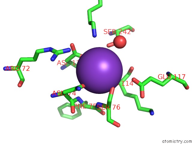
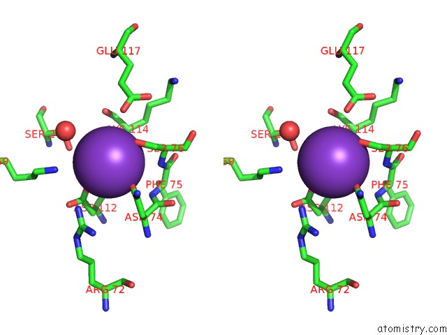
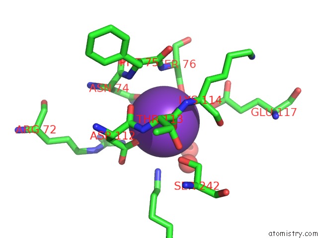
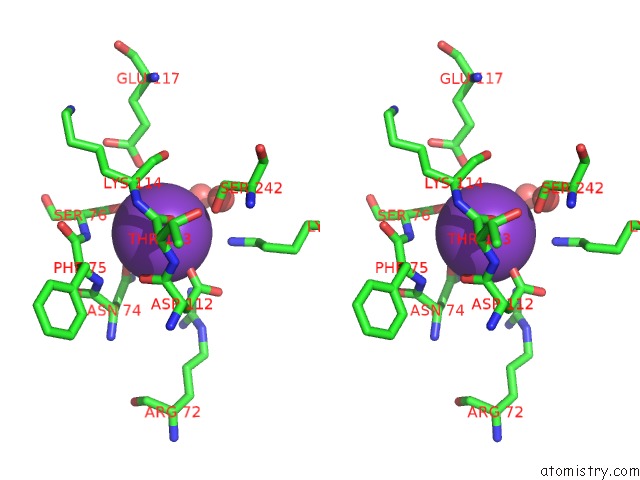
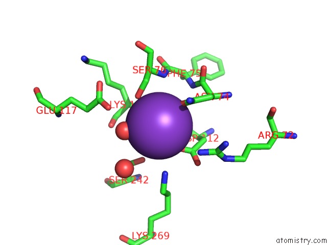
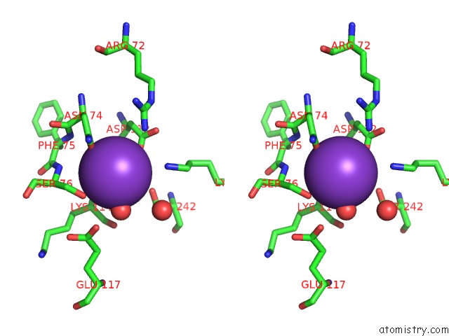
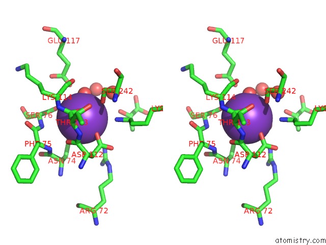
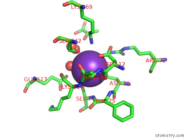
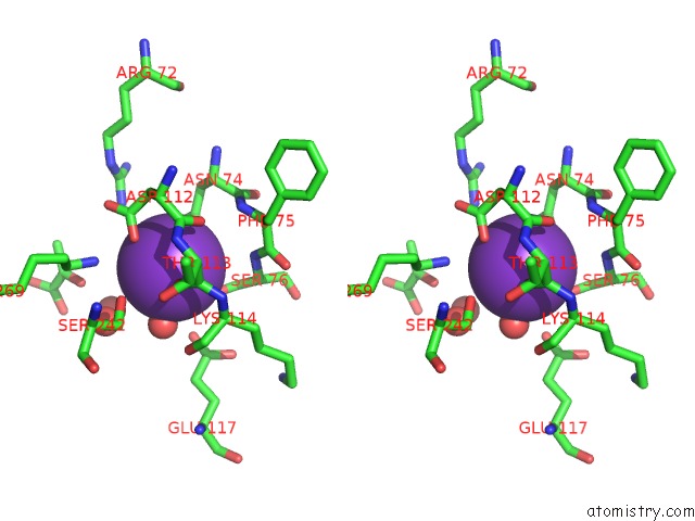
Potassium binding site 1 out of 8 in 3n25
Go back to
Potassium binding site 1 out
of 8 in the The Structure of Muscle Pyruvate Kinase in Complex with Proline, Pyruvate, and MN2+
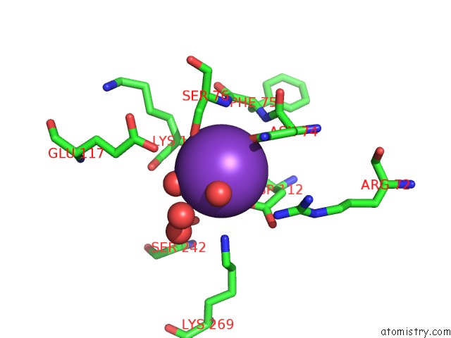
Mono view
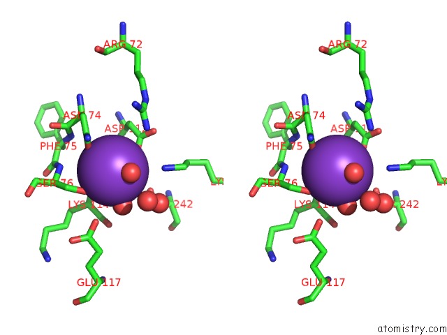
Stereo pair view
Mono view
Stereo pair view
A full contact list of Potassium with other atoms in the K binding
site number 1 of The Structure of Muscle Pyruvate Kinase in Complex with Proline, Pyruvate, and MN2+ within 5.0Å range:
|
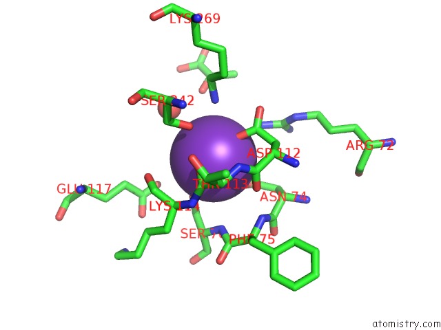
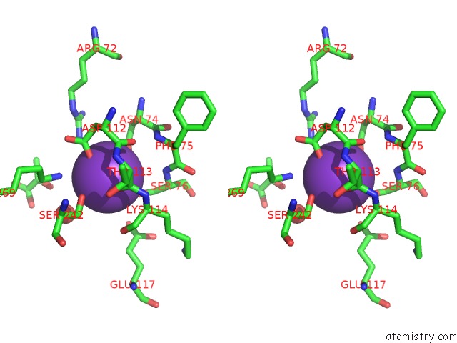
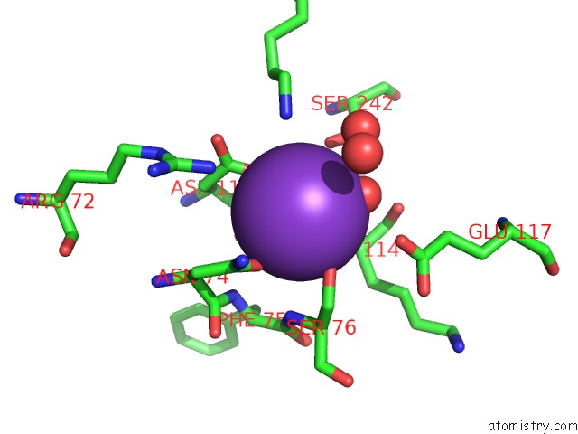
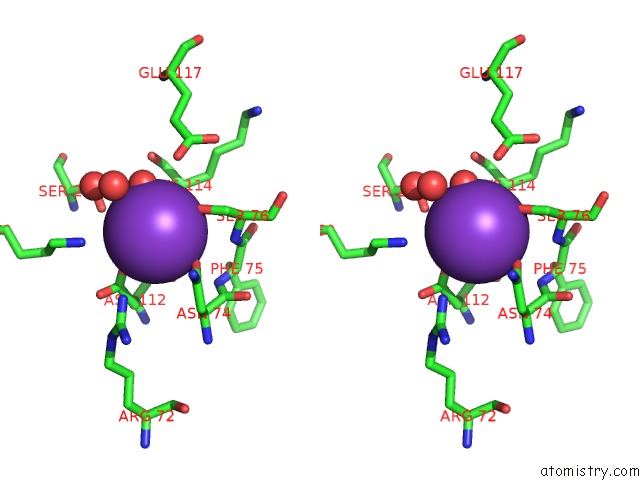
Potassium binding site 2 out of 8 in 3n25
Go back to
Potassium binding site 2 out
of 8 in the The Structure of Muscle Pyruvate Kinase in Complex with Proline, Pyruvate, and MN2+
Mono view
Stereo pair view
Mono view
Stereo pair view
A full contact list of Potassium with other atoms in the K binding
site number 2 of The Structure of Muscle Pyruvate Kinase in Complex with Proline, Pyruvate, and MN2+ within 5.0Å range:
|
Potassium binding site 3 out of 8 in 3n25
Go back to
Potassium binding site 3 out
of 8 in the The Structure of Muscle Pyruvate Kinase in Complex with Proline, Pyruvate, and MN2+
Mono view
Stereo pair view
Mono view
Stereo pair view
A full contact list of Potassium with other atoms in the K binding
site number 3 of The Structure of Muscle Pyruvate Kinase in Complex with Proline, Pyruvate, and MN2+ within 5.0Å range:
|
Potassium binding site 4 out of 8 in 3n25
Go back to
Potassium binding site 4 out
of 8 in the The Structure of Muscle Pyruvate Kinase in Complex with Proline, Pyruvate, and MN2+
Mono view
Stereo pair view
Mono view
Stereo pair view
A full contact list of Potassium with other atoms in the K binding
site number 4 of The Structure of Muscle Pyruvate Kinase in Complex with Proline, Pyruvate, and MN2+ within 5.0Å range:
|
Potassium binding site 5 out of 8 in 3n25
Go back to
Potassium binding site 5 out
of 8 in the The Structure of Muscle Pyruvate Kinase in Complex with Proline, Pyruvate, and MN2+
Mono view
Stereo pair view
Mono view
Stereo pair view
A full contact list of Potassium with other atoms in the K binding
site number 5 of The Structure of Muscle Pyruvate Kinase in Complex with Proline, Pyruvate, and MN2+ within 5.0Å range:
|
Potassium binding site 6 out of 8 in 3n25
Go back to
Potassium binding site 6 out
of 8 in the The Structure of Muscle Pyruvate Kinase in Complex with Proline, Pyruvate, and MN2+
Mono view
Stereo pair view
Mono view
Stereo pair view
A full contact list of Potassium with other atoms in the K binding
site number 6 of The Structure of Muscle Pyruvate Kinase in Complex with Proline, Pyruvate, and MN2+ within 5.0Å range:
|
Potassium binding site 7 out of 8 in 3n25
Go back to
Potassium binding site 7 out
of 8 in the The Structure of Muscle Pyruvate Kinase in Complex with Proline, Pyruvate, and MN2+

Mono view
Stereo pair view
Mono view
Stereo pair view
A full contact list of Potassium with other atoms in the K binding
site number 7 of The Structure of Muscle Pyruvate Kinase in Complex with Proline, Pyruvate, and MN2+ within 5.0Å range:
|
Potassium binding site 8 out of 8 in 3n25
Go back to
Potassium binding site 8 out
of 8 in the The Structure of Muscle Pyruvate Kinase in Complex with Proline, Pyruvate, and MN2+
Mono view
Stereo pair view
Mono view
Stereo pair view
A full contact list of Potassium with other atoms in the K binding
site number 8 of The Structure of Muscle Pyruvate Kinase in Complex with Proline, Pyruvate, and MN2+ within 5.0Å range:
|
Reference:
A.W.Fenton,
T.A.Johnson,
T.Holyoak.
The Pyruvate Kinase Model System, A Cautionary Tale For the Use of Osmolyte Perturbations to Support Conformational Equilibria in Allostery. Protein Sci. V. 19 1796 2010.
ISSN: ISSN 0961-8368
PubMed: 20629175
DOI: 10.1002/PRO.450
Page generated: Mon Aug 12 08:49:00 2024
ISSN: ISSN 0961-8368
PubMed: 20629175
DOI: 10.1002/PRO.450
Last articles
Zn in 9J0NZn in 9J0O
Zn in 9J0P
Zn in 9FJX
Zn in 9EKB
Zn in 9C0F
Zn in 9CAH
Zn in 9CH0
Zn in 9CH3
Zn in 9CH1