Potassium »
PDB 3m62-3ow2 »
3mz4 »
Potassium in PDB 3mz4: Crystal Structure of D101L MN2+ HDAC8 Complexed with M344
Enzymatic activity of Crystal Structure of D101L MN2+ HDAC8 Complexed with M344
All present enzymatic activity of Crystal Structure of D101L MN2+ HDAC8 Complexed with M344:
3.5.1.98;
3.5.1.98;
Protein crystallography data
The structure of Crystal Structure of D101L MN2+ HDAC8 Complexed with M344, PDB code: 3mz4
was solved by
D.P.Dowling,
S.G.Gattis,
C.A.Fierke,
D.W.Christianson,
with X-Ray Crystallography technique. A brief refinement statistics is given in the table below:
| Resolution Low / High (Å) | 45.35 / 1.84 |
| Space group | P 21 21 21 |
| Cell size a, b, c (Å), α, β, γ (°) | 88.353, 91.148, 104.547, 90.00, 90.00, 90.00 |
| R / Rfree (%) | 20.1 / 24.9 |
Other elements in 3mz4:
The structure of Crystal Structure of D101L MN2+ HDAC8 Complexed with M344 also contains other interesting chemical elements:
| Manganese | (Mn) | 2 atoms |
Potassium Binding Sites:
The binding sites of Potassium atom in the Crystal Structure of D101L MN2+ HDAC8 Complexed with M344
(pdb code 3mz4). This binding sites where shown within
5.0 Angstroms radius around Potassium atom.
In total 4 binding sites of Potassium where determined in the Crystal Structure of D101L MN2+ HDAC8 Complexed with M344, PDB code: 3mz4:
Jump to Potassium binding site number: 1; 2; 3; 4;
In total 4 binding sites of Potassium where determined in the Crystal Structure of D101L MN2+ HDAC8 Complexed with M344, PDB code: 3mz4:
Jump to Potassium binding site number: 1; 2; 3; 4;
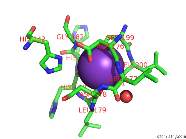
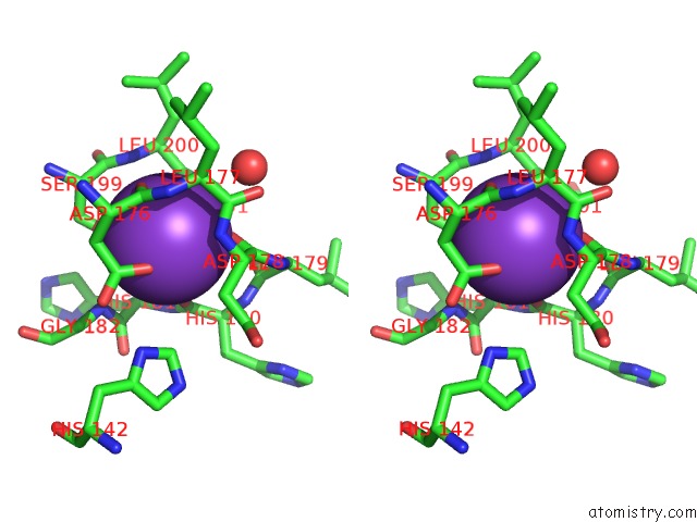
Potassium binding site 1 out of 4 in 3mz4
Go back to
Potassium binding site 1 out
of 4 in the Crystal Structure of D101L MN2+ HDAC8 Complexed with M344
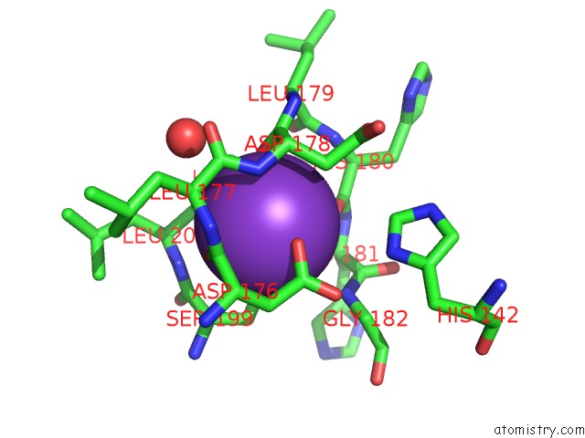
Mono view
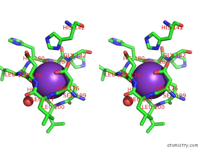
Stereo pair view
Mono view
Stereo pair view
A full contact list of Potassium with other atoms in the K binding
site number 1 of Crystal Structure of D101L MN2+ HDAC8 Complexed with M344 within 5.0Å range:
|
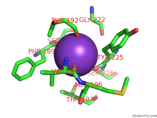
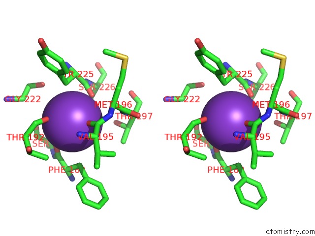
Potassium binding site 2 out of 4 in 3mz4
Go back to
Potassium binding site 2 out
of 4 in the Crystal Structure of D101L MN2+ HDAC8 Complexed with M344
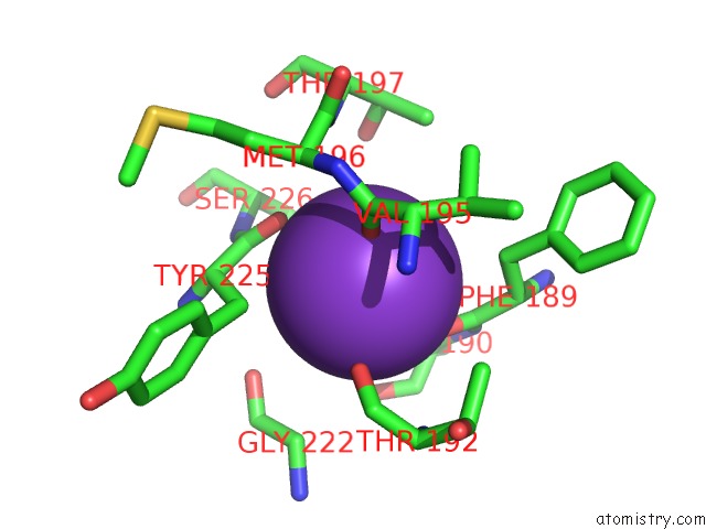
Mono view
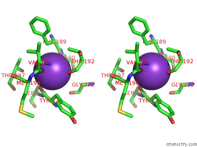
Stereo pair view
Mono view
Stereo pair view
A full contact list of Potassium with other atoms in the K binding
site number 2 of Crystal Structure of D101L MN2+ HDAC8 Complexed with M344 within 5.0Å range:
|
Potassium binding site 3 out of 4 in 3mz4
Go back to
Potassium binding site 3 out
of 4 in the Crystal Structure of D101L MN2+ HDAC8 Complexed with M344
Mono view
Stereo pair view
Mono view
Stereo pair view
A full contact list of Potassium with other atoms in the K binding
site number 3 of Crystal Structure of D101L MN2+ HDAC8 Complexed with M344 within 5.0Å range:
|
Potassium binding site 4 out of 4 in 3mz4
Go back to
Potassium binding site 4 out
of 4 in the Crystal Structure of D101L MN2+ HDAC8 Complexed with M344
Mono view
Stereo pair view
Mono view
Stereo pair view
A full contact list of Potassium with other atoms in the K binding
site number 4 of Crystal Structure of D101L MN2+ HDAC8 Complexed with M344 within 5.0Å range:
|
Reference:
D.P.Dowling,
S.G.Gattis,
C.A.Fierke,
D.W.Christianson.
Structures of Metal-Substituted Human Histone Deacetylase 8 Provide Mechanistic Inferences on Biological Function. Biochemistry V. 49 5048 2010.
ISSN: ISSN 0006-2960
PubMed: 20545365
DOI: 10.1021/BI1005046
Page generated: Mon Aug 12 08:47:33 2024
ISSN: ISSN 0006-2960
PubMed: 20545365
DOI: 10.1021/BI1005046
Last articles
Zn in 9J0NZn in 9J0O
Zn in 9J0P
Zn in 9FJX
Zn in 9EKB
Zn in 9C0F
Zn in 9CAH
Zn in 9CH0
Zn in 9CH3
Zn in 9CH1