Potassium in PDB 3mz3: Crystal Structure of CO2+ HDAC8 Complexed with M344
Enzymatic activity of Crystal Structure of CO2+ HDAC8 Complexed with M344
All present enzymatic activity of Crystal Structure of CO2+ HDAC8 Complexed with M344:
3.5.1.98;
Protein crystallography data
The structure of Crystal Structure of CO2+ HDAC8 Complexed with M344, PDB code: 3mz3
was solved by
D.P.Dowling,
S.G.Gattis,
C.A.Fierke,
D.W.Christianson,
with X-Ray Crystallography technique. A brief refinement statistics is given in the table below:
Resolution Low / High (Å)
|
50.00 /
3.20
|
Space group
|
P 1 21 1
|
Cell size a, b, c (Å), α, β, γ (°)
|
55.626,
86.143,
94.508,
90.00,
94.07,
90.00
|
R / Rfree (%)
|
20.4 /
25.6
|
Other elements in 3mz3:
The structure of Crystal Structure of CO2+ HDAC8 Complexed with M344 also contains other interesting chemical elements:
Potassium Binding Sites:
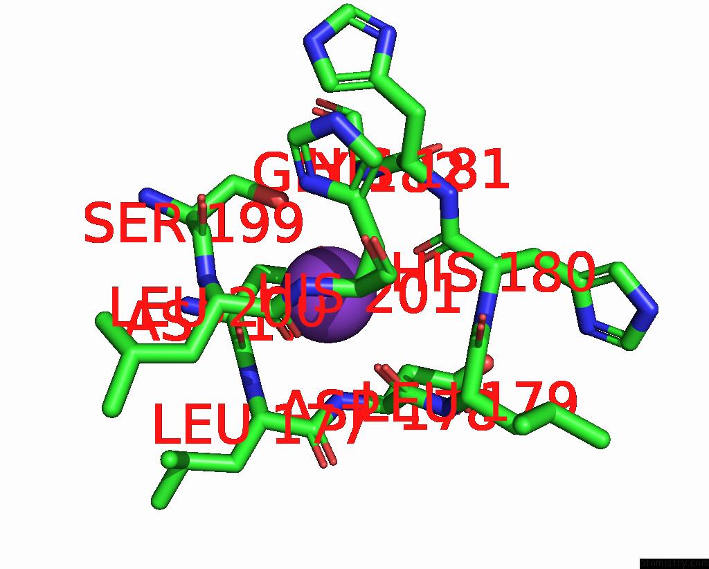
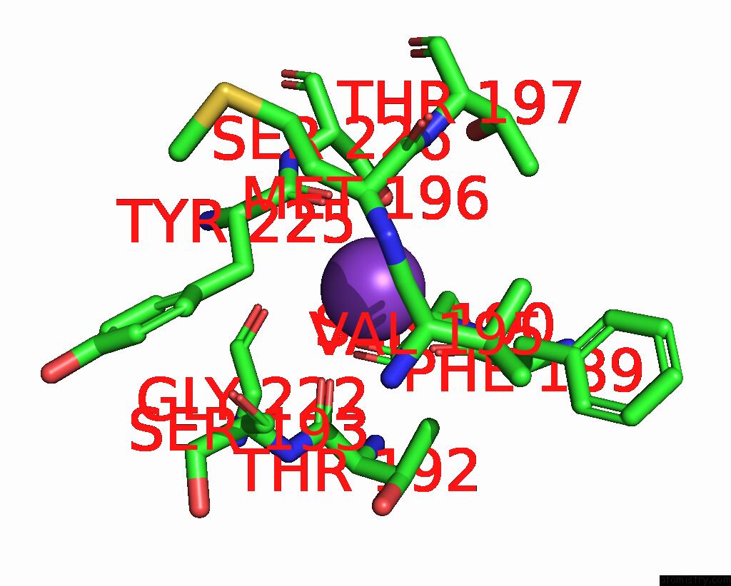
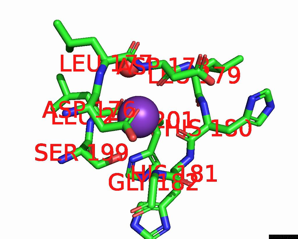
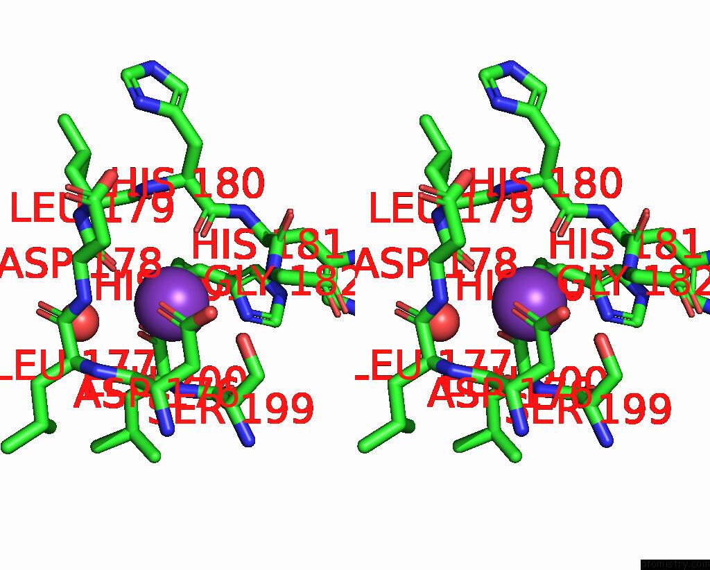
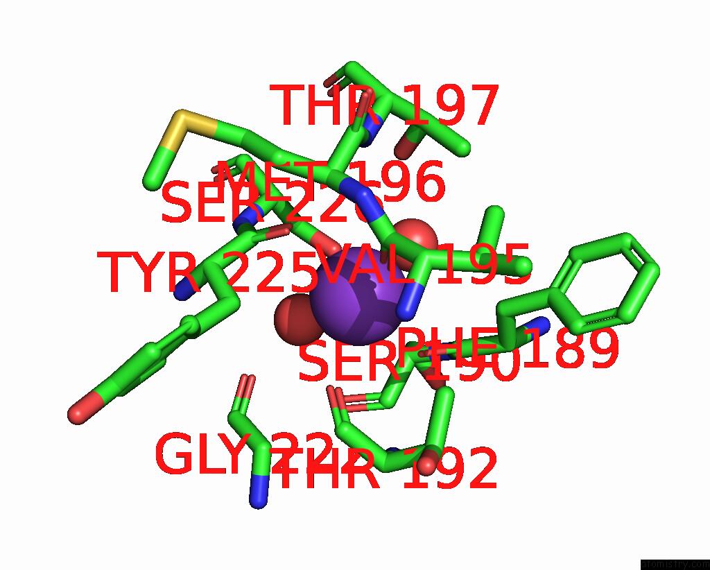
The binding sites of Potassium atom in the Crystal Structure of CO2+ HDAC8 Complexed with M344
(pdb code 3mz3). This binding sites where shown within
5.0 Angstroms radius around Potassium atom.
In total 4 binding sites of Potassium where determined in the
Crystal Structure of CO2+ HDAC8 Complexed with M344, PDB code: 3mz3:
Jump to Potassium binding site number:
1;
2;
3;
4;
Potassium binding site 1 out
of 4 in 3mz3
Go back to
Potassium Binding Sites List in 3mz3
Potassium binding site 1 out
of 4 in the Crystal Structure of CO2+ HDAC8 Complexed with M344
Mono view
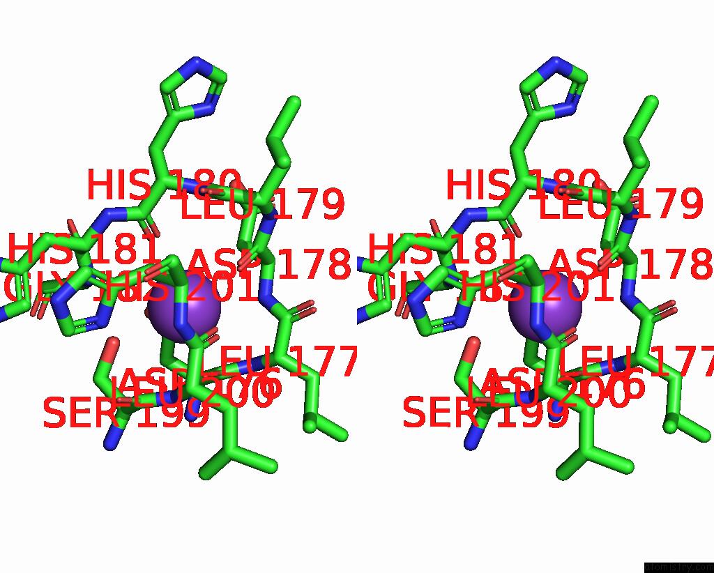
Stereo pair view
|
A full contact list of Potassium with other atoms in the K binding
site number 1 of Crystal Structure of CO2+ HDAC8 Complexed with M344 within 5.0Å range:
probe
|
atom
|
residue
|
distance (Å)
|
B
|
Occ
|
A:K402
b:53.8
occ:1.00
|
OD1
|
A:ASP176
|
2.7
|
63.1
|
1.0
|
O
|
A:ASP178
|
2.7
|
50.7
|
1.0
|
O
|
A:ASP176
|
2.8
|
58.8
|
1.0
|
O
|
A:LEU200
|
2.8
|
57.0
|
1.0
|
O
|
A:HIS180
|
2.9
|
50.4
|
1.0
|
OG
|
A:SER199
|
3.0
|
58.0
|
1.0
|
CG
|
A:ASP176
|
3.3
|
62.9
|
1.0
|
C
|
A:ASP176
|
3.3
|
58.2
|
1.0
|
N
|
A:ASP178
|
3.4
|
53.4
|
1.0
|
CB
|
A:ASP176
|
3.5
|
60.9
|
1.0
|
CB
|
A:HIS201
|
3.6
|
56.9
|
1.0
|
C
|
A:ASP178
|
3.6
|
49.9
|
1.0
|
C
|
A:LEU200
|
3.6
|
56.4
|
1.0
|
C
|
A:HIS180
|
3.9
|
49.0
|
1.0
|
CA
|
A:ASP178
|
3.9
|
51.8
|
1.0
|
N
|
A:LEU177
|
3.9
|
56.9
|
1.0
|
C
|
A:LEU177
|
4.0
|
54.2
|
1.0
|
N
|
A:LEU200
|
4.0
|
54.9
|
1.0
|
CB
|
A:ASP178
|
4.1
|
52.4
|
1.0
|
CA
|
A:ASP176
|
4.1
|
59.6
|
1.0
|
N
|
A:GLY182
|
4.1
|
50.8
|
1.0
|
CA
|
A:LEU177
|
4.1
|
54.9
|
1.0
|
CA
|
A:HIS201
|
4.2
|
56.7
|
1.0
|
CB
|
A:SER199
|
4.2
|
58.8
|
1.0
|
N
|
A:HIS201
|
4.2
|
56.6
|
1.0
|
OD2
|
A:ASP176
|
4.3
|
63.6
|
1.0
|
ND1
|
A:HIS201
|
4.3
|
58.4
|
1.0
|
C
|
A:SER199
|
4.4
|
56.1
|
1.0
|
CG
|
A:HIS201
|
4.4
|
58.1
|
1.0
|
CA
|
A:HIS181
|
4.4
|
49.6
|
1.0
|
CA
|
A:SER199
|
4.4
|
57.7
|
1.0
|
N
|
A:HIS181
|
4.5
|
49.6
|
1.0
|
N
|
A:HIS180
|
4.5
|
47.1
|
1.0
|
CA
|
A:LEU200
|
4.6
|
55.7
|
1.0
|
C
|
A:HIS181
|
4.6
|
50.0
|
1.0
|
N
|
A:LEU179
|
4.7
|
47.6
|
1.0
|
C
|
A:LEU179
|
4.8
|
46.5
|
1.0
|
O
|
A:LEU177
|
4.8
|
53.0
|
1.0
|
CA
|
A:HIS180
|
4.8
|
48.5
|
1.0
|
|
Potassium binding site 2 out
of 4 in 3mz3
Go back to
Potassium Binding Sites List in 3mz3
Potassium binding site 2 out
of 4 in the Crystal Structure of CO2+ HDAC8 Complexed with M344
Mono view
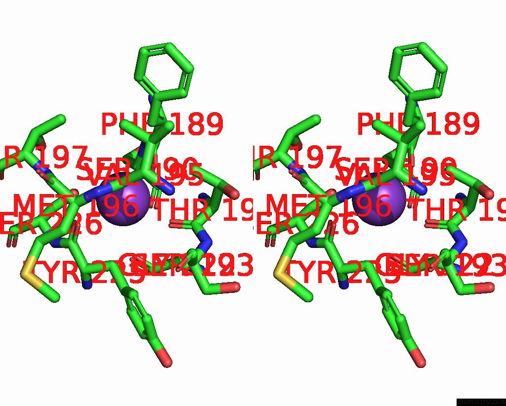
Stereo pair view
|
A full contact list of Potassium with other atoms in the K binding
site number 2 of Crystal Structure of CO2+ HDAC8 Complexed with M344 within 5.0Å range:
probe
|
atom
|
residue
|
distance (Å)
|
B
|
Occ
|
A:K404
b:67.3
occ:1.00
|
O
|
A:PHE189
|
2.6
|
76.2
|
1.0
|
O
|
A:VAL195
|
2.6
|
72.6
|
1.0
|
O
|
A:TYR225
|
2.7
|
83.4
|
1.0
|
O
|
A:THR192
|
2.8
|
82.2
|
1.0
|
CB
|
A:TYR225
|
3.5
|
88.0
|
1.0
|
C
|
A:TYR225
|
3.5
|
83.7
|
1.0
|
C
|
A:PHE189
|
3.6
|
75.3
|
1.0
|
OG
|
A:SER226
|
3.7
|
76.1
|
1.0
|
C
|
A:VAL195
|
3.8
|
74.2
|
1.0
|
C
|
A:THR192
|
3.9
|
81.6
|
1.0
|
CG2
|
A:THR192
|
4.0
|
81.2
|
1.0
|
CA
|
A:TYR225
|
4.1
|
85.8
|
1.0
|
CA
|
A:SER190
|
4.2
|
77.9
|
1.0
|
CA
|
A:MET196
|
4.2
|
73.4
|
1.0
|
O
|
A:GLY222
|
4.2
|
93.6
|
1.0
|
CB
|
A:PHE189
|
4.2
|
72.9
|
1.0
|
N
|
A:SER190
|
4.3
|
76.3
|
1.0
|
N
|
A:SER226
|
4.4
|
81.7
|
1.0
|
C
|
A:SER190
|
4.4
|
79.8
|
1.0
|
O
|
A:SER190
|
4.4
|
81.2
|
1.0
|
N
|
A:THR192
|
4.5
|
81.0
|
1.0
|
N
|
A:MET196
|
4.5
|
73.3
|
1.0
|
N
|
A:THR197
|
4.6
|
69.2
|
1.0
|
CA
|
A:PHE189
|
4.6
|
73.9
|
1.0
|
CA
|
A:THR192
|
4.7
|
81.1
|
1.0
|
CA
|
A:SER226
|
4.7
|
79.1
|
1.0
|
CB
|
A:SER226
|
4.7
|
78.2
|
1.0
|
CG
|
A:TYR225
|
4.8
|
90.2
|
1.0
|
CA
|
A:GLY222
|
4.9
|
93.2
|
1.0
|
C
|
A:MET196
|
4.9
|
71.9
|
1.0
|
N
|
A:SER193
|
4.9
|
81.3
|
1.0
|
N
|
A:TYR225
|
4.9
|
87.4
|
1.0
|
CA
|
A:VAL195
|
4.9
|
75.7
|
1.0
|
|
Potassium binding site 3 out
of 4 in 3mz3
Go back to
Potassium Binding Sites List in 3mz3
Potassium binding site 3 out
of 4 in the Crystal Structure of CO2+ HDAC8 Complexed with M344
Mono view
Stereo pair view
|
A full contact list of Potassium with other atoms in the K binding
site number 3 of Crystal Structure of CO2+ HDAC8 Complexed with M344 within 5.0Å range:
probe
|
atom
|
residue
|
distance (Å)
|
B
|
Occ
|
B:K403
b:53.8
occ:1.00
|
O
|
B:ASP178
|
2.7
|
51.4
|
1.0
|
O
|
B:LEU200
|
2.7
|
58.0
|
1.0
|
OD1
|
B:ASP176
|
2.8
|
57.0
|
1.0
|
O
|
B:ASP176
|
2.9
|
57.2
|
1.0
|
O
|
B:HIS180
|
2.9
|
46.6
|
1.0
|
OG
|
B:SER199
|
3.0
|
57.7
|
1.0
|
CB
|
B:HIS201
|
3.4
|
55.7
|
1.0
|
CG
|
B:ASP176
|
3.4
|
58.0
|
1.0
|
C
|
B:ASP176
|
3.5
|
57.9
|
1.0
|
N
|
B:ASP178
|
3.5
|
54.9
|
1.0
|
C
|
B:LEU200
|
3.5
|
57.6
|
1.0
|
C
|
B:ASP178
|
3.6
|
50.7
|
1.0
|
CB
|
B:ASP176
|
3.7
|
58.7
|
1.0
|
C
|
B:HIS180
|
3.8
|
47.3
|
1.0
|
N
|
B:LEU200
|
4.0
|
57.4
|
1.0
|
CA
|
B:ASP178
|
4.0
|
53.1
|
1.0
|
CA
|
B:HIS201
|
4.0
|
55.1
|
1.0
|
C
|
B:LEU177
|
4.0
|
55.4
|
1.0
|
N
|
B:LEU177
|
4.1
|
56.9
|
1.0
|
N
|
B:HIS201
|
4.1
|
56.6
|
1.0
|
N
|
B:GLY182
|
4.1
|
54.2
|
1.0
|
CB
|
B:SER199
|
4.1
|
60.6
|
1.0
|
CB
|
B:ASP178
|
4.2
|
53.7
|
1.0
|
ND1
|
B:HIS201
|
4.2
|
56.2
|
1.0
|
CA
|
B:ASP176
|
4.2
|
59.0
|
1.0
|
CG
|
B:HIS201
|
4.2
|
56.1
|
1.0
|
CA
|
B:LEU177
|
4.2
|
56.0
|
1.0
|
C
|
B:SER199
|
4.3
|
57.9
|
1.0
|
CA
|
B:HIS181
|
4.3
|
50.0
|
1.0
|
OD2
|
B:ASP176
|
4.4
|
58.7
|
1.0
|
CA
|
B:SER199
|
4.4
|
59.3
|
1.0
|
N
|
B:HIS181
|
4.4
|
48.0
|
1.0
|
N
|
B:HIS180
|
4.5
|
46.0
|
1.0
|
O
|
B:HOH409
|
4.5
|
54.2
|
1.0
|
CA
|
B:LEU200
|
4.5
|
57.4
|
1.0
|
C
|
B:HIS181
|
4.6
|
52.5
|
1.0
|
C
|
B:LEU179
|
4.7
|
44.8
|
1.0
|
N
|
B:LEU179
|
4.7
|
46.8
|
1.0
|
CA
|
B:HIS180
|
4.7
|
46.8
|
1.0
|
O
|
B:LEU177
|
4.9
|
55.0
|
1.0
|
|
Potassium binding site 4 out
of 4 in 3mz3
Go back to
Potassium Binding Sites List in 3mz3
Potassium binding site 4 out
of 4 in the Crystal Structure of CO2+ HDAC8 Complexed with M344
Mono view
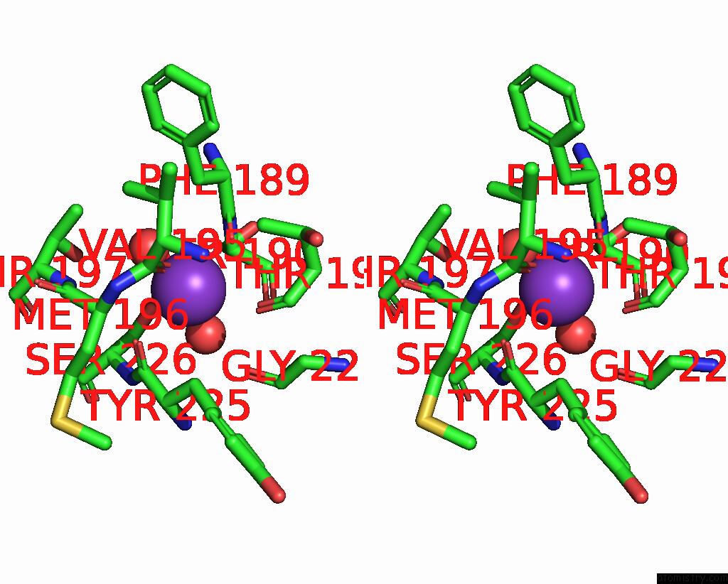
Stereo pair view
|
A full contact list of Potassium with other atoms in the K binding
site number 4 of Crystal Structure of CO2+ HDAC8 Complexed with M344 within 5.0Å range:
probe
|
atom
|
residue
|
distance (Å)
|
B
|
Occ
|
B:K405
b:72.2
occ:1.00
|
O
|
B:PHE189
|
2.5
|
75.4
|
1.0
|
O
|
B:HOH393
|
2.6
|
61.4
|
1.0
|
O
|
B:VAL195
|
2.6
|
72.5
|
1.0
|
O
|
B:TYR225
|
2.7
|
82.2
|
1.0
|
O
|
B:HOH395
|
2.8
|
54.6
|
1.0
|
O
|
B:THR192
|
3.0
|
78.3
|
1.0
|
C
|
B:PHE189
|
3.5
|
74.5
|
1.0
|
C
|
B:TYR225
|
3.5
|
82.7
|
1.0
|
OG
|
B:SER226
|
3.6
|
76.7
|
1.0
|
CB
|
B:TYR225
|
3.7
|
86.5
|
1.0
|
C
|
B:VAL195
|
3.8
|
73.0
|
1.0
|
CA
|
B:SER190
|
4.1
|
76.4
|
1.0
|
CB
|
B:PHE189
|
4.1
|
71.0
|
1.0
|
CG2
|
B:THR192
|
4.1
|
81.6
|
1.0
|
C
|
B:THR192
|
4.1
|
79.2
|
1.0
|
N
|
B:SER190
|
4.2
|
75.7
|
1.0
|
CA
|
B:MET196
|
4.2
|
71.8
|
1.0
|
CA
|
B:TYR225
|
4.2
|
84.5
|
1.0
|
N
|
B:SER226
|
4.3
|
80.7
|
1.0
|
C
|
B:SER190
|
4.4
|
77.2
|
1.0
|
O
|
B:GLY222
|
4.4
|
94.0
|
1.0
|
O
|
B:SER190
|
4.4
|
77.5
|
1.0
|
N
|
B:THR197
|
4.4
|
68.7
|
1.0
|
CA
|
B:PHE189
|
4.5
|
72.7
|
1.0
|
N
|
B:MET196
|
4.5
|
72.0
|
1.0
|
N
|
B:THR192
|
4.6
|
79.5
|
1.0
|
CB
|
B:SER226
|
4.6
|
77.3
|
1.0
|
CA
|
B:SER226
|
4.7
|
78.5
|
1.0
|
CA
|
B:THR192
|
4.8
|
79.7
|
1.0
|
OG1
|
B:THR197
|
4.8
|
63.1
|
1.0
|
C
|
B:MET196
|
4.8
|
71.1
|
1.0
|
CA
|
B:VAL195
|
5.0
|
73.4
|
1.0
|
|
Reference:
D.P.Dowling,
S.G.Gattis,
C.A.Fierke,
D.W.Christianson.
Structures of Metal-Substituted Human Histone Deacetylase 8 Provide Mechanistic Inferences on Biological Function. Biochemistry V. 49 5048 2010.
ISSN: ISSN 0006-2960
PubMed: 20545365
DOI: 10.1021/BI1005046
Page generated: Mon Aug 12 08:47:20 2024
|