Potassium »
PDB 3m62-3ow2 »
3md7 »
Potassium in PDB 3md7: Crystal Structure of A Beta-Lactamase-Like Protein Bound to Gmp From Brucella Melitensis
Protein crystallography data
The structure of Crystal Structure of A Beta-Lactamase-Like Protein Bound to Gmp From Brucella Melitensis, PDB code: 3md7
was solved by
Seattle Structural Genomics Center For Infectious Disease (Ssgcid),
with X-Ray Crystallography technique. A brief refinement statistics is given in the table below:
| Resolution Low / High (Å) | 35.85 / 1.27 |
| Space group | C 2 2 21 |
| Cell size a, b, c (Å), α, β, γ (°) | 72.890, 75.250, 98.360, 90.00, 90.00, 90.00 |
| R / Rfree (%) | 10.7 / 13.4 |
Other elements in 3md7:
The structure of Crystal Structure of A Beta-Lactamase-Like Protein Bound to Gmp From Brucella Melitensis also contains other interesting chemical elements:
| Manganese | (Mn) | 2 atoms |
| Sodium | (Na) | 2 atoms |
Potassium Binding Sites:
The binding sites of Potassium atom in the Crystal Structure of A Beta-Lactamase-Like Protein Bound to Gmp From Brucella Melitensis
(pdb code 3md7). This binding sites where shown within
5.0 Angstroms radius around Potassium atom.
In total only one binding site of Potassium was determined in the Crystal Structure of A Beta-Lactamase-Like Protein Bound to Gmp From Brucella Melitensis, PDB code: 3md7:
In total only one binding site of Potassium was determined in the Crystal Structure of A Beta-Lactamase-Like Protein Bound to Gmp From Brucella Melitensis, PDB code: 3md7:
Potassium binding site 1 out of 1 in 3md7
Go back to
Potassium binding site 1 out
of 1 in the Crystal Structure of A Beta-Lactamase-Like Protein Bound to Gmp From Brucella Melitensis
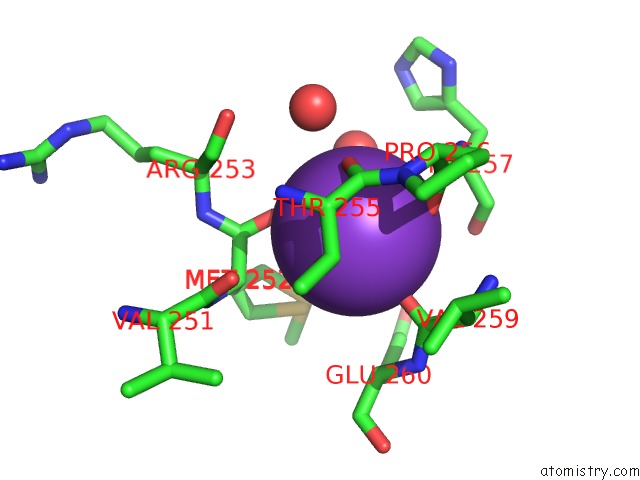
Mono view
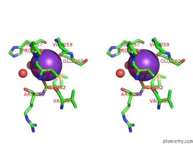
Stereo pair view
Mono view
Stereo pair view
A full contact list of Potassium with other atoms in the K binding
site number 1 of Crystal Structure of A Beta-Lactamase-Like Protein Bound to Gmp From Brucella Melitensis within 5.0Å range:
|
Reference:
J.Abendroth,
B.Sankaran,
T.E.Edwards,
A.S.Gardberg,
S.Dieterich,
J.Bhandari,
A.J.Napuli,
W.C.Van Voorhis,
B.L.Staker,
P.J.Myler,
L.J.Stewart.
Braba.11339.A: Anomalous Diffraction and Ligand Binding Guide Towards the Elucidation of the Function of A `Putative Beta-Lactamase-Like Protein From Brucella Melitensis. Acta Crystallogr.,Sect.F V. 67 1106 2011.
ISSN: ESSN 1744-3091
PubMed: 21904058
DOI: 10.1107/S1744309111010220
Page generated: Mon Aug 12 08:46:48 2024
ISSN: ESSN 1744-3091
PubMed: 21904058
DOI: 10.1107/S1744309111010220
Last articles
Zn in 9J0NZn in 9J0O
Zn in 9J0P
Zn in 9FJX
Zn in 9EKB
Zn in 9C0F
Zn in 9CAH
Zn in 9CH0
Zn in 9CH3
Zn in 9CH1