Potassium »
PDB 3is7-3lut »
3l63 »
Potassium in PDB 3l63: Crystal Structure of Camphor-Bound P450CAM at Low [K+]
Enzymatic activity of Crystal Structure of Camphor-Bound P450CAM at Low [K+]
All present enzymatic activity of Crystal Structure of Camphor-Bound P450CAM at Low [K+]:
1.14.15.1;
1.14.15.1;
Protein crystallography data
The structure of Crystal Structure of Camphor-Bound P450CAM at Low [K+], PDB code: 3l63
was solved by
Y.-T.Lee,
R.F.Wilson,
I.Rupniewski,
D.B.Goodin,
with X-Ray Crystallography technique. A brief refinement statistics is given in the table below:
| Resolution Low / High (Å) | 10.00 / 1.50 |
| Space group | P 1 21 1 |
| Cell size a, b, c (Å), α, β, γ (°) | 35.362, 98.194, 54.197, 90.00, 103.86, 90.00 |
| R / Rfree (%) | 16.4 / 19.7 |
Other elements in 3l63:
The structure of Crystal Structure of Camphor-Bound P450CAM at Low [K+] also contains other interesting chemical elements:
| Iron | (Fe) | 1 atom |
Potassium Binding Sites:
The binding sites of Potassium atom in the Crystal Structure of Camphor-Bound P450CAM at Low [K+]
(pdb code 3l63). This binding sites where shown within
5.0 Angstroms radius around Potassium atom.
In total only one binding site of Potassium was determined in the Crystal Structure of Camphor-Bound P450CAM at Low [K+], PDB code: 3l63:
In total only one binding site of Potassium was determined in the Crystal Structure of Camphor-Bound P450CAM at Low [K+], PDB code: 3l63:
Potassium binding site 1 out of 1 in 3l63
Go back to
Potassium binding site 1 out
of 1 in the Crystal Structure of Camphor-Bound P450CAM at Low [K+]
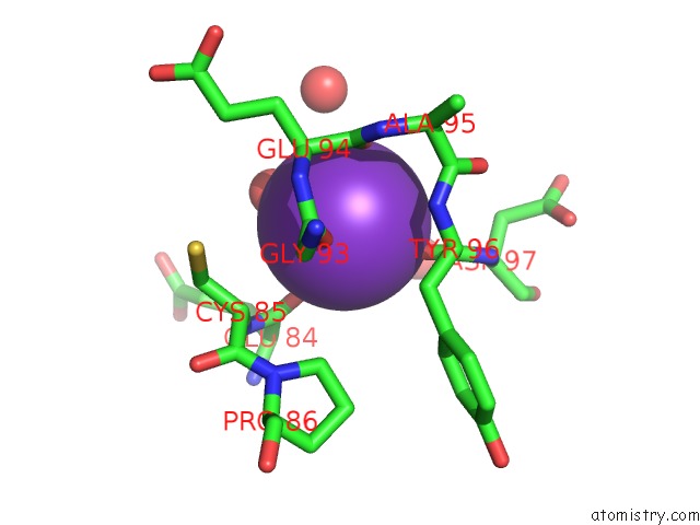
Mono view
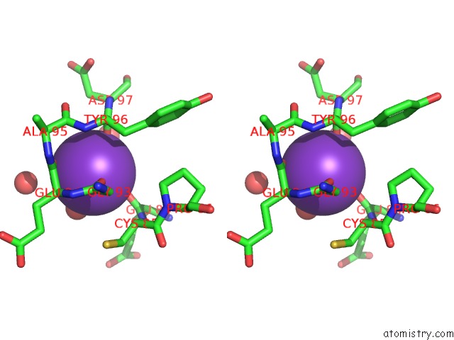
Stereo pair view
Mono view
Stereo pair view
A full contact list of Potassium with other atoms in the K binding
site number 1 of Crystal Structure of Camphor-Bound P450CAM at Low [K+] within 5.0Å range:
|
Reference:
Y.T.Lee,
R.F.Wilson,
I.Rupniewski,
D.B.Goodin.
P450CAM Visits An Open Conformation in the Absence of Substrate. Biochemistry V. 49 3412 2010.
ISSN: ISSN 0006-2960
PubMed: 20297780
DOI: 10.1021/BI100183G
Page generated: Sat Aug 9 05:14:43 2025
ISSN: ISSN 0006-2960
PubMed: 20297780
DOI: 10.1021/BI100183G
Last articles
Mg in 1IJDMg in 1IIR
Mg in 1II9
Mg in 1II6
Mg in 1II0
Mg in 1IHU
Mg in 1IH8
Mg in 1IH1
Mg in 1IGW
Mg in 1IG5