Potassium »
PDB 3is7-3lut »
3l01 »
Potassium in PDB 3l01: Crystal Structure of Monomeric Glycogen Synthase From Pyrococcus Abyssi
Enzymatic activity of Crystal Structure of Monomeric Glycogen Synthase From Pyrococcus Abyssi
All present enzymatic activity of Crystal Structure of Monomeric Glycogen Synthase From Pyrococcus Abyssi:
2.4.1.21;
2.4.1.21;
Protein crystallography data
The structure of Crystal Structure of Monomeric Glycogen Synthase From Pyrococcus Abyssi, PDB code: 3l01
was solved by
A.Diaz,
C.Martinez-Pons,
I.Fita,
J.C.Ferrer,
J.J.Guinovart,
with X-Ray Crystallography technique. A brief refinement statistics is given in the table below:
| Resolution Low / High (Å) | 24.91 / 2.60 |
| Space group | P 21 21 21 |
| Cell size a, b, c (Å), α, β, γ (°) | 103.604, 119.017, 141.101, 90.00, 90.00, 90.00 |
| R / Rfree (%) | 19.8 / 22.8 |
Other elements in 3l01:
The structure of Crystal Structure of Monomeric Glycogen Synthase From Pyrococcus Abyssi also contains other interesting chemical elements:
| Chlorine | (Cl) | 6 atoms |
Potassium Binding Sites:
The binding sites of Potassium atom in the Crystal Structure of Monomeric Glycogen Synthase From Pyrococcus Abyssi
(pdb code 3l01). This binding sites where shown within
5.0 Angstroms radius around Potassium atom.
In total 6 binding sites of Potassium where determined in the Crystal Structure of Monomeric Glycogen Synthase From Pyrococcus Abyssi, PDB code: 3l01:
Jump to Potassium binding site number: 1; 2; 3; 4; 5; 6;
In total 6 binding sites of Potassium where determined in the Crystal Structure of Monomeric Glycogen Synthase From Pyrococcus Abyssi, PDB code: 3l01:
Jump to Potassium binding site number: 1; 2; 3; 4; 5; 6;
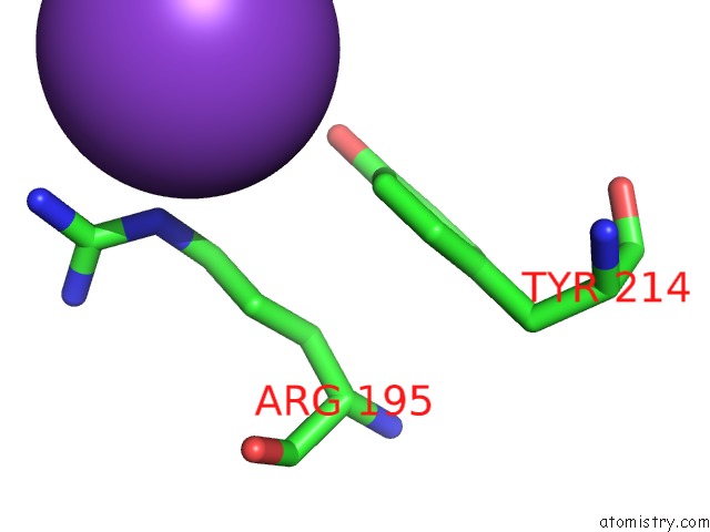
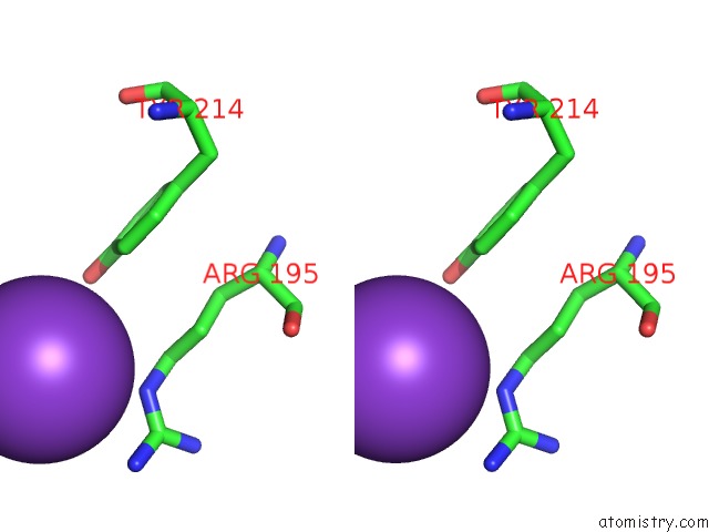
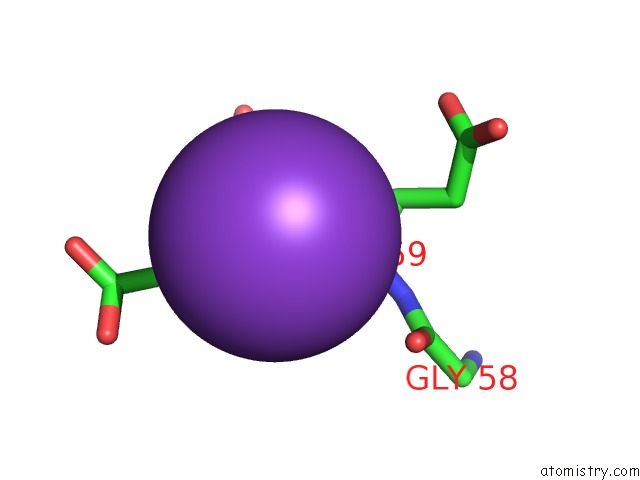
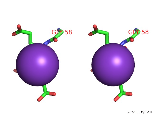
Potassium binding site 1 out of 6 in 3l01
Go back to
Potassium binding site 1 out
of 6 in the Crystal Structure of Monomeric Glycogen Synthase From Pyrococcus Abyssi
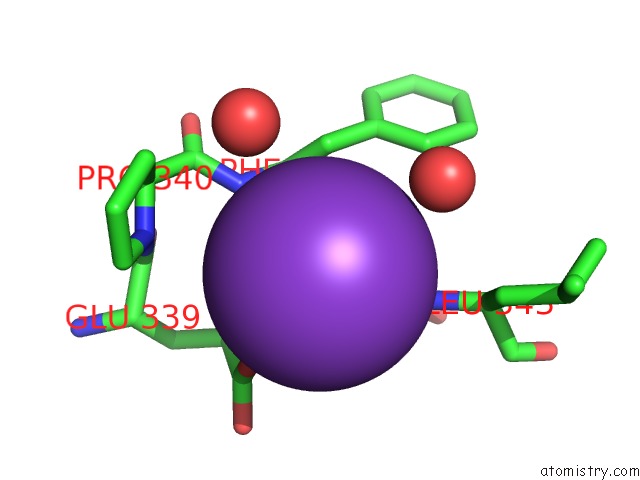
Mono view
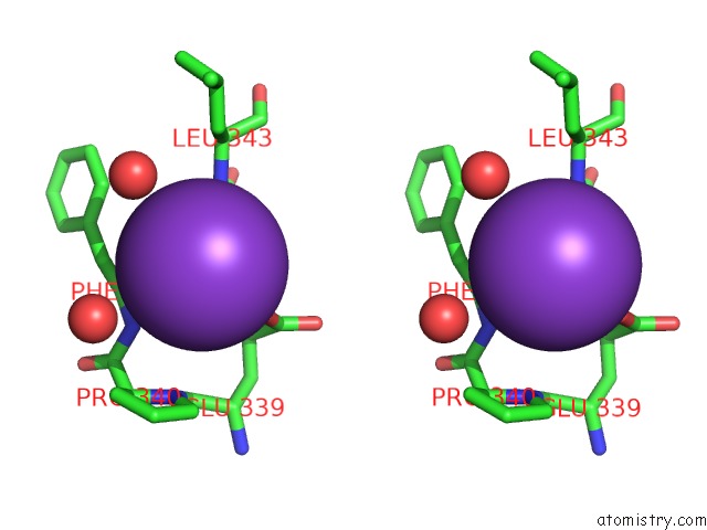
Stereo pair view
Mono view
Stereo pair view
A full contact list of Potassium with other atoms in the K binding
site number 1 of Crystal Structure of Monomeric Glycogen Synthase From Pyrococcus Abyssi within 5.0Å range:
|
Potassium binding site 2 out of 6 in 3l01
Go back to
Potassium binding site 2 out
of 6 in the Crystal Structure of Monomeric Glycogen Synthase From Pyrococcus Abyssi
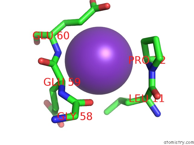
Mono view
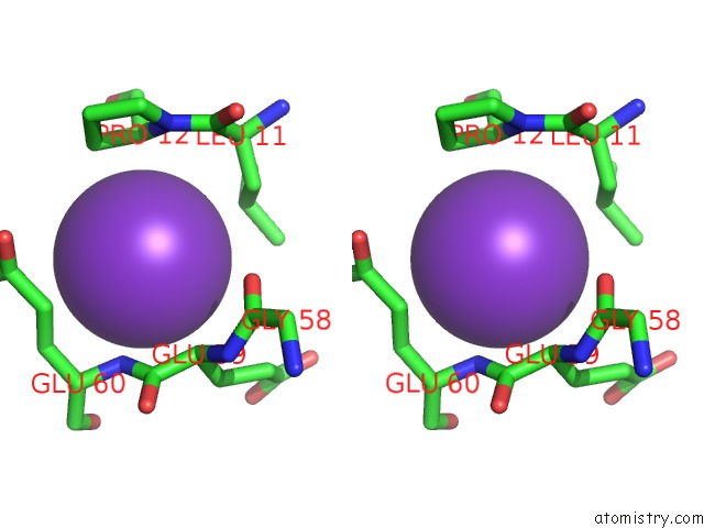
Stereo pair view
Mono view
Stereo pair view
A full contact list of Potassium with other atoms in the K binding
site number 2 of Crystal Structure of Monomeric Glycogen Synthase From Pyrococcus Abyssi within 5.0Å range:
|
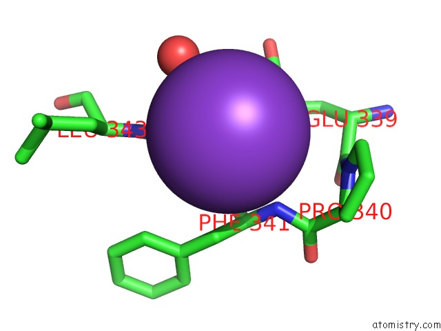
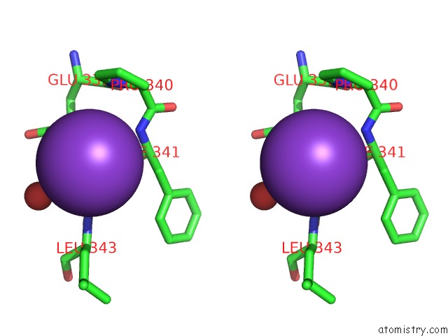
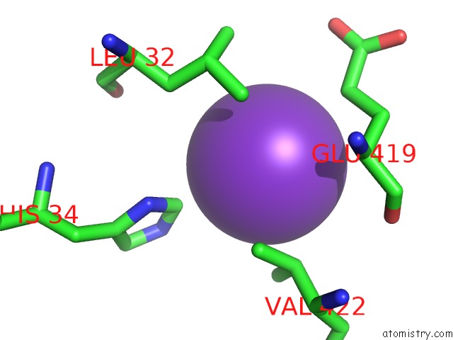
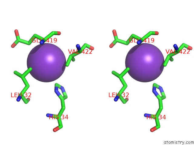
Potassium binding site 3 out of 6 in 3l01
Go back to
Potassium binding site 3 out
of 6 in the Crystal Structure of Monomeric Glycogen Synthase From Pyrococcus Abyssi
Mono view
Stereo pair view
Mono view
Stereo pair view
A full contact list of Potassium with other atoms in the K binding
site number 3 of Crystal Structure of Monomeric Glycogen Synthase From Pyrococcus Abyssi within 5.0Å range:
|
Potassium binding site 4 out of 6 in 3l01
Go back to
Potassium binding site 4 out
of 6 in the Crystal Structure of Monomeric Glycogen Synthase From Pyrococcus Abyssi
Mono view
Stereo pair view
Mono view
Stereo pair view
A full contact list of Potassium with other atoms in the K binding
site number 4 of Crystal Structure of Monomeric Glycogen Synthase From Pyrococcus Abyssi within 5.0Å range:
|
Potassium binding site 5 out of 6 in 3l01
Go back to
Potassium binding site 5 out
of 6 in the Crystal Structure of Monomeric Glycogen Synthase From Pyrococcus Abyssi
Mono view
Stereo pair view
Mono view
Stereo pair view
A full contact list of Potassium with other atoms in the K binding
site number 5 of Crystal Structure of Monomeric Glycogen Synthase From Pyrococcus Abyssi within 5.0Å range:
|
Potassium binding site 6 out of 6 in 3l01
Go back to
Potassium binding site 6 out
of 6 in the Crystal Structure of Monomeric Glycogen Synthase From Pyrococcus Abyssi
Mono view
Stereo pair view
Mono view
Stereo pair view
A full contact list of Potassium with other atoms in the K binding
site number 6 of Crystal Structure of Monomeric Glycogen Synthase From Pyrococcus Abyssi within 5.0Å range:
|
Reference:
A.Diaz,
C.Martinez-Pons,
I.Fita,
J.C.Ferrer,
J.J.Guinovart.
Processivity and Subcellular Localization of Glycogen Synthase Depend on A Non-Catalytic High Affinity Glycogen-Binding Site. J.Biol.Chem. V. 286 18505 2011.
ISSN: ISSN 0021-9258
PubMed: 21464127
DOI: 10.1074/JBC.M111.236109
Page generated: Sat Aug 9 05:14:10 2025
ISSN: ISSN 0021-9258
PubMed: 21464127
DOI: 10.1074/JBC.M111.236109
Last articles
K in 6B2WK in 6B10
K in 6AI6
K in 6AU4
K in 6ASO
K in 6AFZ
K in 6AFY
K in 6AFX
K in 6AFW
K in 6AFV