Potassium »
PDB 3is7-3lut »
3ise »
Potassium in PDB 3ise: Structure of Mineralized Bfrb (Double Soak) From Pseudomonas Aeruginosa to 2.8A Resolution
Protein crystallography data
The structure of Structure of Mineralized Bfrb (Double Soak) From Pseudomonas Aeruginosa to 2.8A Resolution, PDB code: 3ise
was solved by
S.Lovell,
S.K.Weeratunga,
K.P.Battaile,
M.Rivera,
with X-Ray Crystallography technique. A brief refinement statistics is given in the table below:
| Resolution Low / High (Å) | 47.51 / 2.80 |
| Space group | P 21 21 21 |
| Cell size a, b, c (Å), α, β, γ (°) | 125.712, 203.206, 207.865, 90.00, 90.00, 90.00 |
| R / Rfree (%) | 19.5 / 23.7 |
Other elements in 3ise:
The structure of Structure of Mineralized Bfrb (Double Soak) From Pseudomonas Aeruginosa to 2.8A Resolution also contains other interesting chemical elements:
| Iron | (Fe) | 36 atoms |
Potassium Binding Sites:
The binding sites of Potassium atom in the Structure of Mineralized Bfrb (Double Soak) From Pseudomonas Aeruginosa to 2.8A Resolution
(pdb code 3ise). This binding sites where shown within
5.0 Angstroms radius around Potassium atom.
In total 6 binding sites of Potassium where determined in the Structure of Mineralized Bfrb (Double Soak) From Pseudomonas Aeruginosa to 2.8A Resolution, PDB code: 3ise:
Jump to Potassium binding site number: 1; 2; 3; 4; 5; 6;
In total 6 binding sites of Potassium where determined in the Structure of Mineralized Bfrb (Double Soak) From Pseudomonas Aeruginosa to 2.8A Resolution, PDB code: 3ise:
Jump to Potassium binding site number: 1; 2; 3; 4; 5; 6;
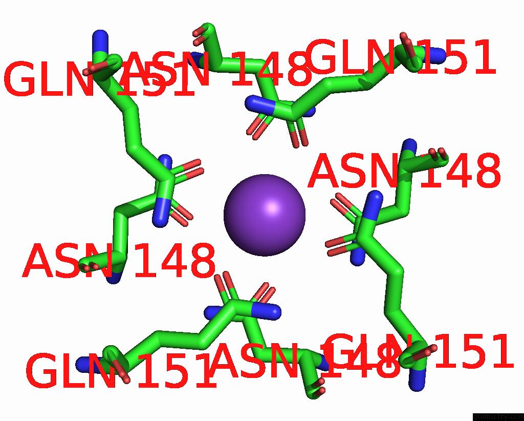
Potassium binding site 1 out of 6 in 3ise
Go back to
Potassium binding site 1 out
of 6 in the Structure of Mineralized Bfrb (Double Soak) From Pseudomonas Aeruginosa to 2.8A Resolution

Mono view

Stereo pair view
Mono view
Stereo pair view
A full contact list of Potassium with other atoms in the K binding
site number 1 of Structure of Mineralized Bfrb (Double Soak) From Pseudomonas Aeruginosa to 2.8A Resolution within 5.0Å range:
|
Potassium binding site 2 out of 6 in 3ise
Go back to
Potassium binding site 2 out
of 6 in the Structure of Mineralized Bfrb (Double Soak) From Pseudomonas Aeruginosa to 2.8A Resolution

Mono view

Stereo pair view
Mono view
Stereo pair view
A full contact list of Potassium with other atoms in the K binding
site number 2 of Structure of Mineralized Bfrb (Double Soak) From Pseudomonas Aeruginosa to 2.8A Resolution within 5.0Å range:
|
Potassium binding site 3 out of 6 in 3ise
Go back to
Potassium binding site 3 out
of 6 in the Structure of Mineralized Bfrb (Double Soak) From Pseudomonas Aeruginosa to 2.8A Resolution

Mono view

Stereo pair view
Mono view
Stereo pair view
A full contact list of Potassium with other atoms in the K binding
site number 3 of Structure of Mineralized Bfrb (Double Soak) From Pseudomonas Aeruginosa to 2.8A Resolution within 5.0Å range:
|
Potassium binding site 4 out of 6 in 3ise
Go back to
Potassium binding site 4 out
of 6 in the Structure of Mineralized Bfrb (Double Soak) From Pseudomonas Aeruginosa to 2.8A Resolution

Mono view

Stereo pair view
Mono view
Stereo pair view
A full contact list of Potassium with other atoms in the K binding
site number 4 of Structure of Mineralized Bfrb (Double Soak) From Pseudomonas Aeruginosa to 2.8A Resolution within 5.0Å range:
|
Potassium binding site 5 out of 6 in 3ise
Go back to
Potassium binding site 5 out
of 6 in the Structure of Mineralized Bfrb (Double Soak) From Pseudomonas Aeruginosa to 2.8A Resolution
Mono view

Stereo pair view
Mono view
Stereo pair view
A full contact list of Potassium with other atoms in the K binding
site number 5 of Structure of Mineralized Bfrb (Double Soak) From Pseudomonas Aeruginosa to 2.8A Resolution within 5.0Å range:
|
Potassium binding site 6 out of 6 in 3ise
Go back to
Potassium binding site 6 out
of 6 in the Structure of Mineralized Bfrb (Double Soak) From Pseudomonas Aeruginosa to 2.8A Resolution

Mono view

Stereo pair view
Mono view
Stereo pair view
A full contact list of Potassium with other atoms in the K binding
site number 6 of Structure of Mineralized Bfrb (Double Soak) From Pseudomonas Aeruginosa to 2.8A Resolution within 5.0Å range:
|
Reference:
S.K.Weeratunga,
S.Lovell,
H.Yao,
K.P.Battaile,
C.J.Fischer,
C.E.Gee,
M.Rivera.
Structural Studies of Bacterioferritin B From Pseudomonas Aeruginosa Suggest A Gating Mechanism For Iron Uptake Via the Ferroxidase Center Biochemistry V. 49 1160 2010.
ISSN: ISSN 0006-2960
PubMed: 20067302
DOI: 10.1021/BI9015204
Page generated: Mon Aug 12 08:37:58 2024
ISSN: ISSN 0006-2960
PubMed: 20067302
DOI: 10.1021/BI9015204
Last articles
Zn in 9JYWZn in 9IR4
Zn in 9IR3
Zn in 9GMX
Zn in 9GMW
Zn in 9JEJ
Zn in 9ERF
Zn in 9ERE
Zn in 9EGV
Zn in 9EGW