Potassium »
PDB 3gvx-3irp »
3irp »
Potassium in PDB 3irp: Crystal Structure of Functional Region of Uafa From Staphylococcus Saprophyticus at 1.50 Angstrom Resolution
Protein crystallography data
The structure of Crystal Structure of Functional Region of Uafa From Staphylococcus Saprophyticus at 1.50 Angstrom Resolution, PDB code: 3irp
was solved by
Y.Tanaka,
Y.Shouji,
E.Matsuoka,
M.Kuroda,
I.Tanaka,
M.Yao,
with X-Ray Crystallography technique. A brief refinement statistics is given in the table below:
| Resolution Low / High (Å) | 19.18 / 1.50 |
| Space group | P 1 21 1 |
| Cell size a, b, c (Å), α, β, γ (°) | 37.935, 126.926, 45.830, 90.00, 96.75, 90.00 |
| R / Rfree (%) | 16.5 / 18.6 |
Potassium Binding Sites:
The binding sites of Potassium atom in the Crystal Structure of Functional Region of Uafa From Staphylococcus Saprophyticus at 1.50 Angstrom Resolution
(pdb code 3irp). This binding sites where shown within
5.0 Angstroms radius around Potassium atom.
In total 5 binding sites of Potassium where determined in the Crystal Structure of Functional Region of Uafa From Staphylococcus Saprophyticus at 1.50 Angstrom Resolution, PDB code: 3irp:
Jump to Potassium binding site number: 1; 2; 3; 4; 5;
In total 5 binding sites of Potassium where determined in the Crystal Structure of Functional Region of Uafa From Staphylococcus Saprophyticus at 1.50 Angstrom Resolution, PDB code: 3irp:
Jump to Potassium binding site number: 1; 2; 3; 4; 5;
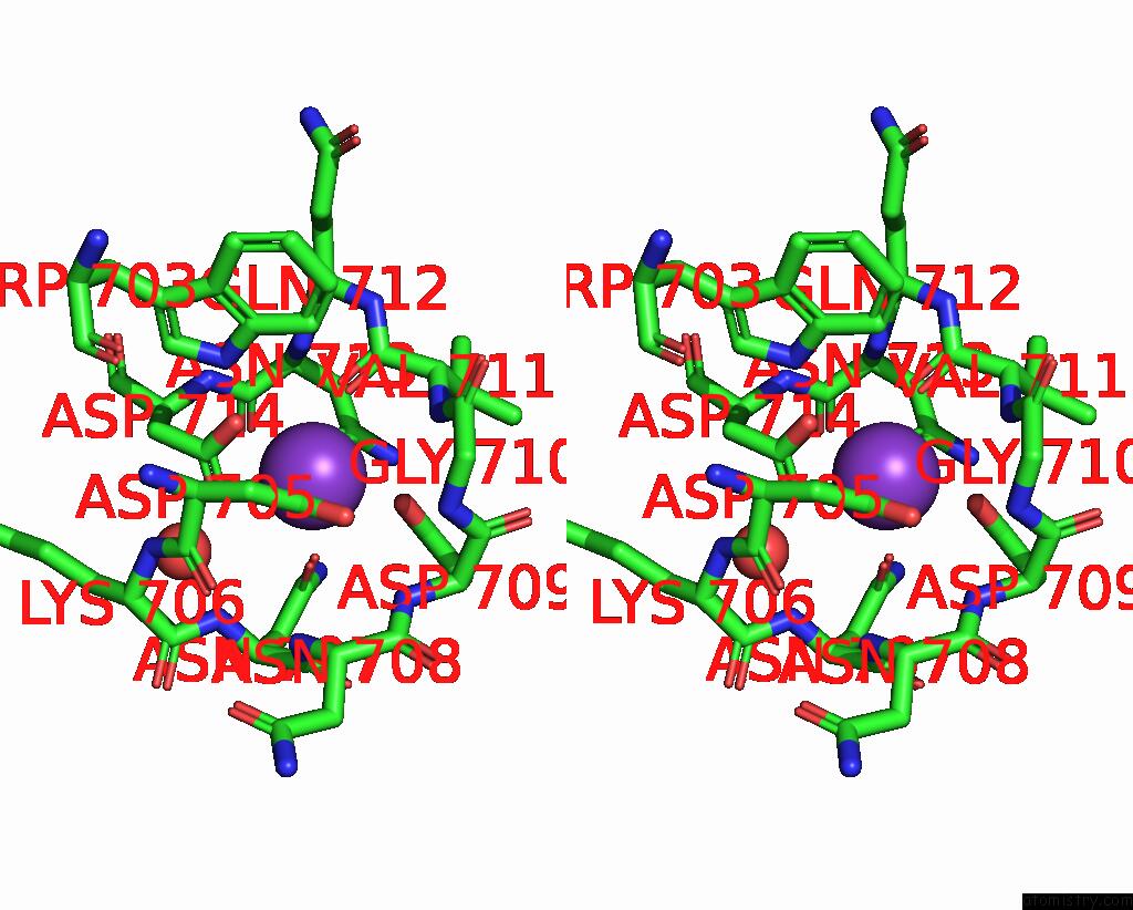
Potassium binding site 1 out of 5 in 3irp
Go back to
Potassium binding site 1 out
of 5 in the Crystal Structure of Functional Region of Uafa From Staphylococcus Saprophyticus at 1.50 Angstrom Resolution

Mono view

Stereo pair view
Mono view
Stereo pair view
A full contact list of Potassium with other atoms in the K binding
site number 1 of Crystal Structure of Functional Region of Uafa From Staphylococcus Saprophyticus at 1.50 Angstrom Resolution within 5.0Å range:
|
Potassium binding site 2 out of 5 in 3irp
Go back to
Potassium binding site 2 out
of 5 in the Crystal Structure of Functional Region of Uafa From Staphylococcus Saprophyticus at 1.50 Angstrom Resolution

Mono view

Stereo pair view
Mono view
Stereo pair view
A full contact list of Potassium with other atoms in the K binding
site number 2 of Crystal Structure of Functional Region of Uafa From Staphylococcus Saprophyticus at 1.50 Angstrom Resolution within 5.0Å range:
|
Potassium binding site 3 out of 5 in 3irp
Go back to
Potassium binding site 3 out
of 5 in the Crystal Structure of Functional Region of Uafa From Staphylococcus Saprophyticus at 1.50 Angstrom Resolution

Mono view
Stereo pair view
Mono view
Stereo pair view
A full contact list of Potassium with other atoms in the K binding
site number 3 of Crystal Structure of Functional Region of Uafa From Staphylococcus Saprophyticus at 1.50 Angstrom Resolution within 5.0Å range:
|
Potassium binding site 4 out of 5 in 3irp
Go back to
Potassium binding site 4 out
of 5 in the Crystal Structure of Functional Region of Uafa From Staphylococcus Saprophyticus at 1.50 Angstrom Resolution

Mono view

Stereo pair view
Mono view
Stereo pair view
A full contact list of Potassium with other atoms in the K binding
site number 4 of Crystal Structure of Functional Region of Uafa From Staphylococcus Saprophyticus at 1.50 Angstrom Resolution within 5.0Å range:
|
Potassium binding site 5 out of 5 in 3irp
Go back to
Potassium binding site 5 out
of 5 in the Crystal Structure of Functional Region of Uafa From Staphylococcus Saprophyticus at 1.50 Angstrom Resolution

Mono view

Stereo pair view
Mono view
Stereo pair view
A full contact list of Potassium with other atoms in the K binding
site number 5 of Crystal Structure of Functional Region of Uafa From Staphylococcus Saprophyticus at 1.50 Angstrom Resolution within 5.0Å range:
|
Reference:
E.Matsuoka,
Y.Tanaka,
M.Kuroda,
Y.Shouji,
T.Ohta,
I.Tanaka,
M.Yao.
Crystal Structure of the Functional Region of Uro-Adherence Factor A From Staphylococcus Saprophyticus Reveals Participation of the B Domain in Ligand Binding Protein Sci. V. 20 406 2011.
ISSN: ISSN 0961-8368
PubMed: 21280131
DOI: 10.1002/PRO.573
Page generated: Mon Aug 12 08:34:30 2024
ISSN: ISSN 0961-8368
PubMed: 21280131
DOI: 10.1002/PRO.573
Last articles
Zn in 9J0NZn in 9J0O
Zn in 9J0P
Zn in 9FJX
Zn in 9EKB
Zn in 9C0F
Zn in 9CAH
Zn in 9CH0
Zn in 9CH3
Zn in 9CH1