Potassium »
PDB 3f2y-3gvf »
3fwp »
Potassium in PDB 3fwp: X-Ray Structure of Uridine Nucleoside Phosphorylease From Salmonella Typhimurium Complexed with Phosphate and Its Inhibitor 2,2'- Anhydrouridine at 1.86 A Resolution
Enzymatic activity of X-Ray Structure of Uridine Nucleoside Phosphorylease From Salmonella Typhimurium Complexed with Phosphate and Its Inhibitor 2,2'- Anhydrouridine at 1.86 A Resolution
All present enzymatic activity of X-Ray Structure of Uridine Nucleoside Phosphorylease From Salmonella Typhimurium Complexed with Phosphate and Its Inhibitor 2,2'- Anhydrouridine at 1.86 A Resolution:
2.4.2.3;
2.4.2.3;
Protein crystallography data
The structure of X-Ray Structure of Uridine Nucleoside Phosphorylease From Salmonella Typhimurium Complexed with Phosphate and Its Inhibitor 2,2'- Anhydrouridine at 1.86 A Resolution, PDB code: 3fwp
was solved by
S.A.Lashkov,
A.M.Mikhailov,
A.G.Gabdulkhakov,
with X-Ray Crystallography technique. A brief refinement statistics is given in the table below:
| Resolution Low / High (Å) | 27.99 / 1.86 |
| Space group | P 21 21 21 |
| Cell size a, b, c (Å), α, β, γ (°) | 88.790, 124.070, 134.100, 90.00, 90.00, 90.00 |
| R / Rfree (%) | 17.6 / 20.6 |
Potassium Binding Sites:
The binding sites of Potassium atom in the X-Ray Structure of Uridine Nucleoside Phosphorylease From Salmonella Typhimurium Complexed with Phosphate and Its Inhibitor 2,2'- Anhydrouridine at 1.86 A Resolution
(pdb code 3fwp). This binding sites where shown within
5.0 Angstroms radius around Potassium atom.
In total 3 binding sites of Potassium where determined in the X-Ray Structure of Uridine Nucleoside Phosphorylease From Salmonella Typhimurium Complexed with Phosphate and Its Inhibitor 2,2'- Anhydrouridine at 1.86 A Resolution, PDB code: 3fwp:
Jump to Potassium binding site number: 1; 2; 3;
In total 3 binding sites of Potassium where determined in the X-Ray Structure of Uridine Nucleoside Phosphorylease From Salmonella Typhimurium Complexed with Phosphate and Its Inhibitor 2,2'- Anhydrouridine at 1.86 A Resolution, PDB code: 3fwp:
Jump to Potassium binding site number: 1; 2; 3;
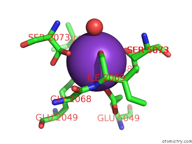
Potassium binding site 1 out of 3 in 3fwp
Go back to
Potassium binding site 1 out
of 3 in the X-Ray Structure of Uridine Nucleoside Phosphorylease From Salmonella Typhimurium Complexed with Phosphate and Its Inhibitor 2,2'- Anhydrouridine at 1.86 A Resolution

Mono view

Stereo pair view
Mono view
Stereo pair view
A full contact list of Potassium with other atoms in the K binding
site number 1 of X-Ray Structure of Uridine Nucleoside Phosphorylease From Salmonella Typhimurium Complexed with Phosphate and Its Inhibitor 2,2'- Anhydrouridine at 1.86 A Resolution within 5.0Å range:
|
Potassium binding site 2 out of 3 in 3fwp
Go back to
Potassium binding site 2 out
of 3 in the X-Ray Structure of Uridine Nucleoside Phosphorylease From Salmonella Typhimurium Complexed with Phosphate and Its Inhibitor 2,2'- Anhydrouridine at 1.86 A Resolution
Mono view

Stereo pair view
Mono view
Stereo pair view
A full contact list of Potassium with other atoms in the K binding
site number 2 of X-Ray Structure of Uridine Nucleoside Phosphorylease From Salmonella Typhimurium Complexed with Phosphate and Its Inhibitor 2,2'- Anhydrouridine at 1.86 A Resolution within 5.0Å range:
|
Potassium binding site 3 out of 3 in 3fwp
Go back to
Potassium binding site 3 out
of 3 in the X-Ray Structure of Uridine Nucleoside Phosphorylease From Salmonella Typhimurium Complexed with Phosphate and Its Inhibitor 2,2'- Anhydrouridine at 1.86 A Resolution

Mono view

Stereo pair view
Mono view
Stereo pair view
A full contact list of Potassium with other atoms in the K binding
site number 3 of X-Ray Structure of Uridine Nucleoside Phosphorylease From Salmonella Typhimurium Complexed with Phosphate and Its Inhibitor 2,2'- Anhydrouridine at 1.86 A Resolution within 5.0Å range:
|
Reference:
A.A.Lashkov,
N.E.Zhukhlistova,
A.H.Gabdoulkhakov,
A.A.Shtil,
R.G.Efremov,
C.Betzel,
A.M.Mikhailov.
The X-Ray Structure of Salmonella Typhimurium Uridine Nucleoside Phosphorylase Complexed with 2,2'-Anhydrouridine, Phosphate and Potassium Ions at 1.86 A Resolution. Acta Crystallogr.,Sect.D V. 66 51 2010.
ISSN: ISSN 0907-4449
PubMed: 20057049
DOI: 10.1107/S0907444909044175
Page generated: Mon Aug 12 08:20:50 2024
ISSN: ISSN 0907-4449
PubMed: 20057049
DOI: 10.1107/S0907444909044175
Last articles
Zn in 9J0NZn in 9J0O
Zn in 9J0P
Zn in 9FJX
Zn in 9EKB
Zn in 9C0F
Zn in 9CAH
Zn in 9CH0
Zn in 9CH3
Zn in 9CH1