Potassium »
PDB 3du0-3f2t »
3e9d »
Potassium in PDB 3e9d: Structure of Full-Length Tigar From Danio Rerio
Protein crystallography data
The structure of Structure of Full-Length Tigar From Danio Rerio, PDB code: 3e9d
was solved by
H.Li,
G.Jogl,
with X-Ray Crystallography technique. A brief refinement statistics is given in the table below:
| Resolution Low / High (Å) | 30.00 / 2.00 |
| Space group | C 1 2 1 |
| Cell size a, b, c (Å), α, β, γ (°) | 127.661, 115.541, 61.311, 90.00, 104.66, 90.00 |
| R / Rfree (%) | 17.1 / 21.3 |
Potassium Binding Sites:
The binding sites of Potassium atom in the Structure of Full-Length Tigar From Danio Rerio
(pdb code 3e9d). This binding sites where shown within
5.0 Angstroms radius around Potassium atom.
In total 2 binding sites of Potassium where determined in the Structure of Full-Length Tigar From Danio Rerio, PDB code: 3e9d:
Jump to Potassium binding site number: 1; 2;
In total 2 binding sites of Potassium where determined in the Structure of Full-Length Tigar From Danio Rerio, PDB code: 3e9d:
Jump to Potassium binding site number: 1; 2;
Potassium binding site 1 out of 2 in 3e9d
Go back to
Potassium binding site 1 out
of 2 in the Structure of Full-Length Tigar From Danio Rerio
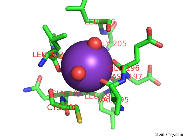
Mono view
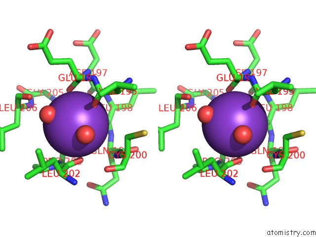
Stereo pair view
Mono view
Stereo pair view
A full contact list of Potassium with other atoms in the K binding
site number 1 of Structure of Full-Length Tigar From Danio Rerio within 5.0Å range:
|
Potassium binding site 2 out of 2 in 3e9d
Go back to
Potassium binding site 2 out
of 2 in the Structure of Full-Length Tigar From Danio Rerio
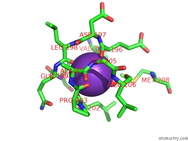
Mono view
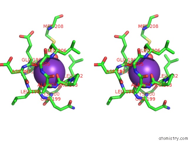
Stereo pair view
Mono view
Stereo pair view
A full contact list of Potassium with other atoms in the K binding
site number 2 of Structure of Full-Length Tigar From Danio Rerio within 5.0Å range:
|
Reference:
H.Li,
G.Jogl.
Tigar (TP53 Induced Glycolysis and Apoptosis Regulator) Is A Fructose-2,6- and Fructose-1,6-Bisphosphatase To Be Published.
Page generated: Mon Aug 12 08:05:05 2024
Last articles
Zn in 9JYWZn in 9IR4
Zn in 9IR3
Zn in 9GMX
Zn in 9GMW
Zn in 9JEJ
Zn in 9ERF
Zn in 9ERE
Zn in 9EGV
Zn in 9EGW