Potassium »
PDB 3du0-3f2t »
3du0 »
Potassium in PDB 3du0: E. Coli Dihydrodipicolinate Synthase with First Substrate, Pyruvate, Bound in Active Site
Enzymatic activity of E. Coli Dihydrodipicolinate Synthase with First Substrate, Pyruvate, Bound in Active Site
All present enzymatic activity of E. Coli Dihydrodipicolinate Synthase with First Substrate, Pyruvate, Bound in Active Site:
4.2.1.52;
4.2.1.52;
Protein crystallography data
The structure of E. Coli Dihydrodipicolinate Synthase with First Substrate, Pyruvate, Bound in Active Site, PDB code: 3du0
was solved by
R.C.J.Dobson,
S.R.A.Devenish,
J.A.Gerrard,
G.B.Jameson,
with X-Ray Crystallography technique. A brief refinement statistics is given in the table below:
| Resolution Low / High (Å) | 32.14 / 2.00 |
| Space group | P 31 2 1 |
| Cell size a, b, c (Å), α, β, γ (°) | 120.950, 120.950, 110.030, 90.00, 90.00, 120.00 |
| R / Rfree (%) | 18.8 / 24.5 |
Other elements in 3du0:
The structure of E. Coli Dihydrodipicolinate Synthase with First Substrate, Pyruvate, Bound in Active Site also contains other interesting chemical elements:
| Chlorine | (Cl) | 4 atoms |
Potassium Binding Sites:
The binding sites of Potassium atom in the E. Coli Dihydrodipicolinate Synthase with First Substrate, Pyruvate, Bound in Active Site
(pdb code 3du0). This binding sites where shown within
5.0 Angstroms radius around Potassium atom.
In total 4 binding sites of Potassium where determined in the E. Coli Dihydrodipicolinate Synthase with First Substrate, Pyruvate, Bound in Active Site, PDB code: 3du0:
Jump to Potassium binding site number: 1; 2; 3; 4;
In total 4 binding sites of Potassium where determined in the E. Coli Dihydrodipicolinate Synthase with First Substrate, Pyruvate, Bound in Active Site, PDB code: 3du0:
Jump to Potassium binding site number: 1; 2; 3; 4;
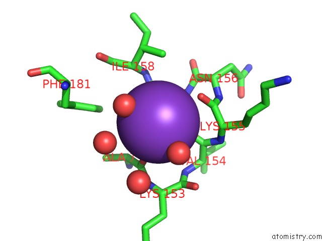
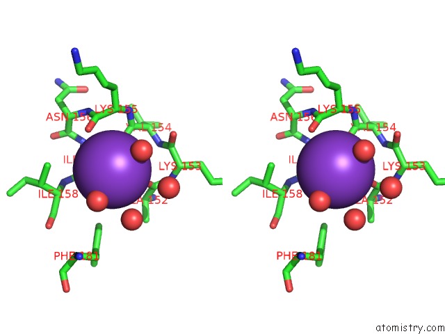
Potassium binding site 1 out of 4 in 3du0
Go back to
Potassium binding site 1 out
of 4 in the E. Coli Dihydrodipicolinate Synthase with First Substrate, Pyruvate, Bound in Active Site
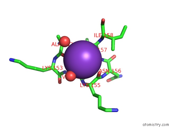
Mono view
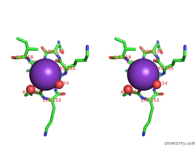
Stereo pair view
Mono view
Stereo pair view
A full contact list of Potassium with other atoms in the K binding
site number 1 of E. Coli Dihydrodipicolinate Synthase with First Substrate, Pyruvate, Bound in Active Site within 5.0Å range:
|
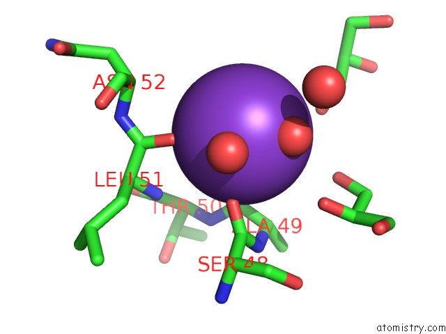
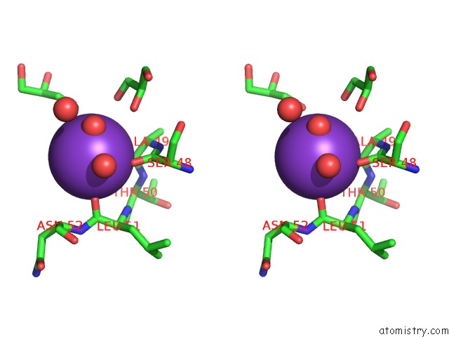
Potassium binding site 2 out of 4 in 3du0
Go back to
Potassium binding site 2 out
of 4 in the E. Coli Dihydrodipicolinate Synthase with First Substrate, Pyruvate, Bound in Active Site
Mono view
Stereo pair view
Mono view
Stereo pair view
A full contact list of Potassium with other atoms in the K binding
site number 2 of E. Coli Dihydrodipicolinate Synthase with First Substrate, Pyruvate, Bound in Active Site within 5.0Å range:
|
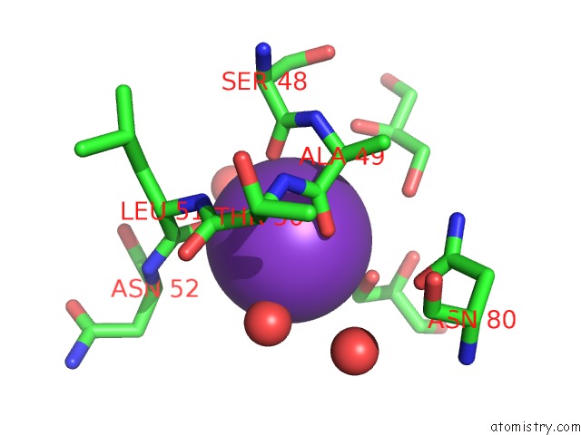
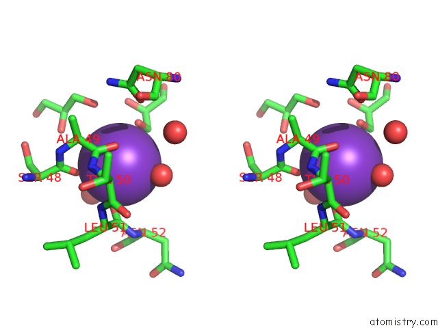
Potassium binding site 3 out of 4 in 3du0
Go back to
Potassium binding site 3 out
of 4 in the E. Coli Dihydrodipicolinate Synthase with First Substrate, Pyruvate, Bound in Active Site
Mono view
Stereo pair view
Mono view
Stereo pair view
A full contact list of Potassium with other atoms in the K binding
site number 3 of E. Coli Dihydrodipicolinate Synthase with First Substrate, Pyruvate, Bound in Active Site within 5.0Å range:
|
Potassium binding site 4 out of 4 in 3du0
Go back to
Potassium binding site 4 out
of 4 in the E. Coli Dihydrodipicolinate Synthase with First Substrate, Pyruvate, Bound in Active Site
Mono view
Stereo pair view
Mono view
Stereo pair view
A full contact list of Potassium with other atoms in the K binding
site number 4 of E. Coli Dihydrodipicolinate Synthase with First Substrate, Pyruvate, Bound in Active Site within 5.0Å range:
|
Reference:
S.R.Devenish,
J.A.Gerrard,
G.B.Jameson,
R.C.Dobson.
The High-Resolution Structure of Dihydrodipicolinate Synthase From Escherichia Coli Bound to Its First Substrate, Pyruvate. Acta Crystallogr.,Sect.F V. 64 1092 2008.
ISSN: ESSN 1744-3091
PubMed: 19052357
DOI: 10.1107/S1744309108033654
Page generated: Mon Aug 12 08:02:54 2024
ISSN: ESSN 1744-3091
PubMed: 19052357
DOI: 10.1107/S1744309108033654
Last articles
Zn in 9MJ5Zn in 9HNW
Zn in 9G0L
Zn in 9FNE
Zn in 9DZN
Zn in 9E0I
Zn in 9D32
Zn in 9DAK
Zn in 8ZXC
Zn in 8ZUF