Potassium »
PDB 3ay9-3ccl »
3bwm »
Potassium in PDB 3bwm: Crystal Structure of Human Catechol O-Methyltransferase with Bound Sam and Dnc
Enzymatic activity of Crystal Structure of Human Catechol O-Methyltransferase with Bound Sam and Dnc
All present enzymatic activity of Crystal Structure of Human Catechol O-Methyltransferase with Bound Sam and Dnc:
2.1.1.6;
2.1.1.6;
Protein crystallography data
The structure of Crystal Structure of Human Catechol O-Methyltransferase with Bound Sam and Dnc, PDB code: 3bwm
was solved by
K.Rutherford,
I.Le Trong,
R.E.Stenkamp,
W.W.Parson,
with X-Ray Crystallography technique. A brief refinement statistics is given in the table below:
| Resolution Low / High (Å) | 33.31 / 1.98 |
| Space group | P 21 21 21 |
| Cell size a, b, c (Å), α, β, γ (°) | 43.284, 66.648, 68.046, 90.00, 90.00, 90.00 |
| R / Rfree (%) | 17.5 / 22.5 |
Other elements in 3bwm:
The structure of Crystal Structure of Human Catechol O-Methyltransferase with Bound Sam and Dnc also contains other interesting chemical elements:
| Magnesium | (Mg) | 1 atom |
Potassium Binding Sites:
The binding sites of Potassium atom in the Crystal Structure of Human Catechol O-Methyltransferase with Bound Sam and Dnc
(pdb code 3bwm). This binding sites where shown within
5.0 Angstroms radius around Potassium atom.
In total only one binding site of Potassium was determined in the Crystal Structure of Human Catechol O-Methyltransferase with Bound Sam and Dnc, PDB code: 3bwm:
In total only one binding site of Potassium was determined in the Crystal Structure of Human Catechol O-Methyltransferase with Bound Sam and Dnc, PDB code: 3bwm:
Potassium binding site 1 out of 1 in 3bwm
Go back to
Potassium binding site 1 out
of 1 in the Crystal Structure of Human Catechol O-Methyltransferase with Bound Sam and Dnc

Mono view
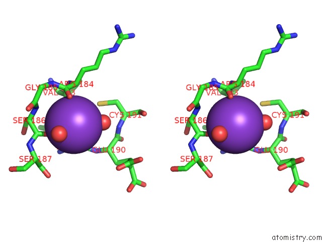
Stereo pair view
Mono view
Stereo pair view
A full contact list of Potassium with other atoms in the K binding
site number 1 of Crystal Structure of Human Catechol O-Methyltransferase with Bound Sam and Dnc within 5.0Å range:
|
Reference:
K.Rutherford,
I.Le Trong,
R.E.Stenkamp,
W.W.Parson.
Crystal Structures of Human 108V and 108M Catechol O-Methyltransferase. J.Mol.Biol. V. 380 120 2008.
ISSN: ISSN 0022-2836
PubMed: 18486144
DOI: 10.1016/J.JMB.2008.04.040
Page generated: Mon Aug 12 07:51:46 2024
ISSN: ISSN 0022-2836
PubMed: 18486144
DOI: 10.1016/J.JMB.2008.04.040
Last articles
Zn in 9J0NZn in 9J0O
Zn in 9J0P
Zn in 9FJX
Zn in 9EKB
Zn in 9C0F
Zn in 9CAH
Zn in 9CH0
Zn in 9CH3
Zn in 9CH1