Potassium »
PDB 2xo1-3atv »
2zuh »
Potassium in PDB 2zuh: Crystal Structure of Camphor-Soaked Ferric Cytochrome P450CAM Mutant (D297A)
Enzymatic activity of Crystal Structure of Camphor-Soaked Ferric Cytochrome P450CAM Mutant (D297A)
All present enzymatic activity of Crystal Structure of Camphor-Soaked Ferric Cytochrome P450CAM Mutant (D297A):
1.14.15.1;
1.14.15.1;
Protein crystallography data
The structure of Crystal Structure of Camphor-Soaked Ferric Cytochrome P450CAM Mutant (D297A), PDB code: 2zuh
was solved by
K.Sakurai,
K.Harada,
H.Shimada,
K.Shimokata,
T.Hayashi,
T.Tsukihara,
with X-Ray Crystallography technique. A brief refinement statistics is given in the table below:
| Resolution Low / High (Å) | 33.54 / 1.55 |
| Space group | P 43 21 2 |
| Cell size a, b, c (Å), α, β, γ (°) | 63.749, 63.749, 250.943, 90.00, 90.00, 90.00 |
| R / Rfree (%) | 16 / 18.5 |
Other elements in 2zuh:
The structure of Crystal Structure of Camphor-Soaked Ferric Cytochrome P450CAM Mutant (D297A) also contains other interesting chemical elements:
| Iron | (Fe) | 1 atom |
Potassium Binding Sites:
The binding sites of Potassium atom in the Crystal Structure of Camphor-Soaked Ferric Cytochrome P450CAM Mutant (D297A)
(pdb code 2zuh). This binding sites where shown within
5.0 Angstroms radius around Potassium atom.
In total only one binding site of Potassium was determined in the Crystal Structure of Camphor-Soaked Ferric Cytochrome P450CAM Mutant (D297A), PDB code: 2zuh:
In total only one binding site of Potassium was determined in the Crystal Structure of Camphor-Soaked Ferric Cytochrome P450CAM Mutant (D297A), PDB code: 2zuh:
Potassium binding site 1 out of 1 in 2zuh
Go back to
Potassium binding site 1 out
of 1 in the Crystal Structure of Camphor-Soaked Ferric Cytochrome P450CAM Mutant (D297A)
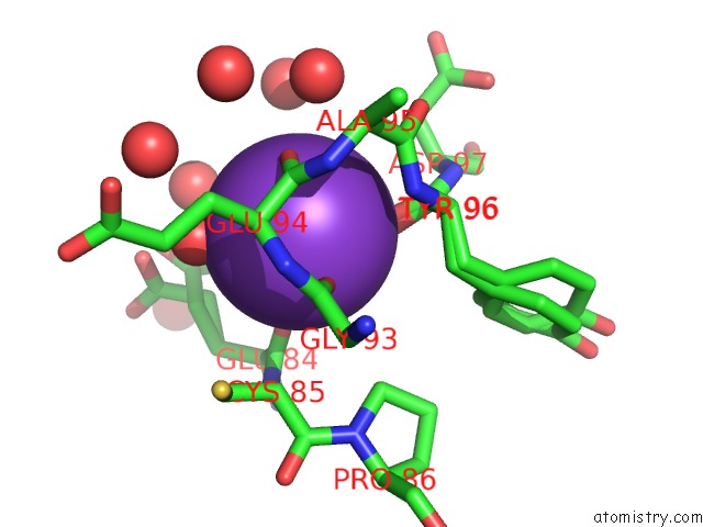
Mono view
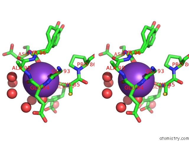
Stereo pair view
Mono view
Stereo pair view
A full contact list of Potassium with other atoms in the K binding
site number 1 of Crystal Structure of Camphor-Soaked Ferric Cytochrome P450CAM Mutant (D297A) within 5.0Å range:
|
Reference:
K.Sakurai,
K.Harada,
H.Shimada,
K.Shimokata,
T.Hayashi,
T.Tsukihara.
Crystal Structure of Camphor-Soaked Ferric Cytochrome P450CAM Mutant To Be Published.
Page generated: Sat Aug 9 04:29:20 2025
Last articles
Mg in 1HJ6Mg in 1HBN
Mg in 1HBU
Mg in 1HH4
Mg in 1HE8
Mg in 1HE1
Mg in 1HDI
Mg in 1HCK
Mg in 1HBO
Mg in 1HBM