Potassium »
PDB 2w0f-2xo0 »
2xdr »
Potassium in PDB 2xdr: Crystallographic Structure of Betaine Aldehyde Dehydrogenase Mutant E252A From Pseudomonas Aeruginosa
Enzymatic activity of Crystallographic Structure of Betaine Aldehyde Dehydrogenase Mutant E252A From Pseudomonas Aeruginosa
All present enzymatic activity of Crystallographic Structure of Betaine Aldehyde Dehydrogenase Mutant E252A From Pseudomonas Aeruginosa:
1.2.1.8;
1.2.1.8;
Protein crystallography data
The structure of Crystallographic Structure of Betaine Aldehyde Dehydrogenase Mutant E252A From Pseudomonas Aeruginosa, PDB code: 2xdr
was solved by
A.G.Diaz-Sanchez,
L.Gonzalez-Segura,
E.Rudino-Pinera,
A.Lira-Rocha,
R.A.Munoz-Clares,
with X-Ray Crystallography technique. A brief refinement statistics is given in the table below:
| Resolution Low / High (Å) | 29.881 / 2.30 |
| Space group | P 32 2 1 |
| Cell size a, b, c (Å), α, β, γ (°) | 151.555, 151.555, 242.406, 90.00, 90.00, 120.00 |
| R / Rfree (%) | 16.03 / 19.97 |
Potassium Binding Sites:
The binding sites of Potassium atom in the Crystallographic Structure of Betaine Aldehyde Dehydrogenase Mutant E252A From Pseudomonas Aeruginosa
(pdb code 2xdr). This binding sites where shown within
5.0 Angstroms radius around Potassium atom.
In total 8 binding sites of Potassium where determined in the Crystallographic Structure of Betaine Aldehyde Dehydrogenase Mutant E252A From Pseudomonas Aeruginosa, PDB code: 2xdr:
Jump to Potassium binding site number: 1; 2; 3; 4; 5; 6; 7; 8;
In total 8 binding sites of Potassium where determined in the Crystallographic Structure of Betaine Aldehyde Dehydrogenase Mutant E252A From Pseudomonas Aeruginosa, PDB code: 2xdr:
Jump to Potassium binding site number: 1; 2; 3; 4; 5; 6; 7; 8;
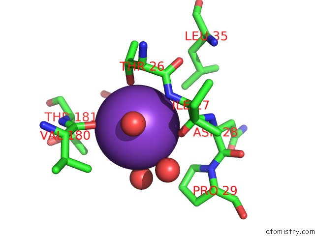
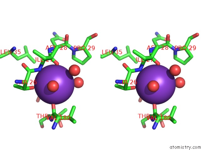
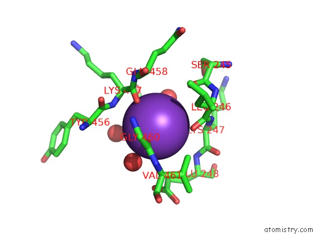
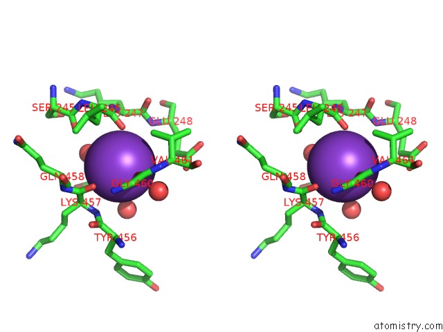
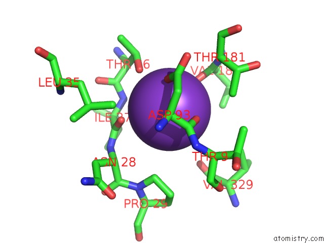
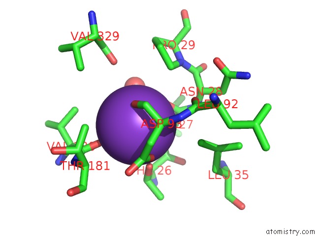
Potassium binding site 1 out of 8 in 2xdr
Go back to
Potassium binding site 1 out
of 8 in the Crystallographic Structure of Betaine Aldehyde Dehydrogenase Mutant E252A From Pseudomonas Aeruginosa
Mono view
Stereo pair view
Mono view
Stereo pair view
A full contact list of Potassium with other atoms in the K binding
site number 1 of Crystallographic Structure of Betaine Aldehyde Dehydrogenase Mutant E252A From Pseudomonas Aeruginosa within 5.0Å range:
|
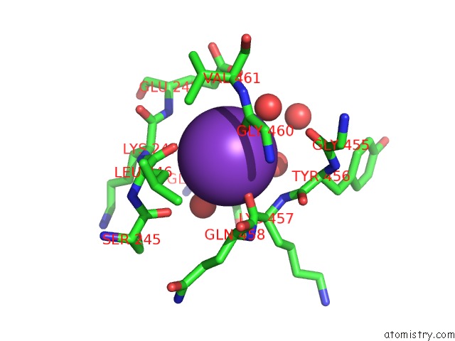
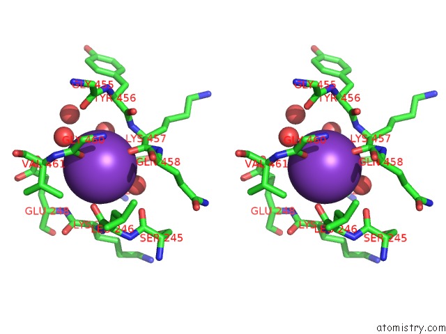
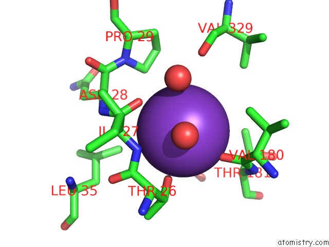
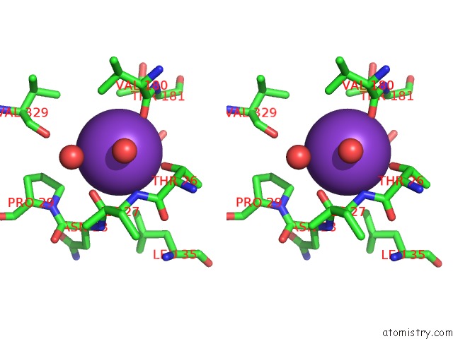
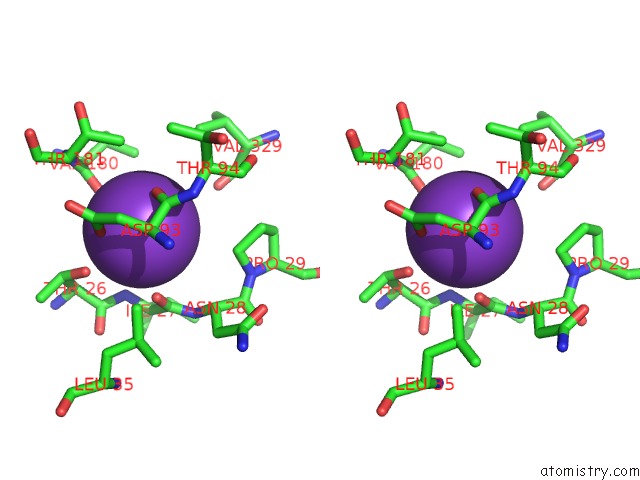
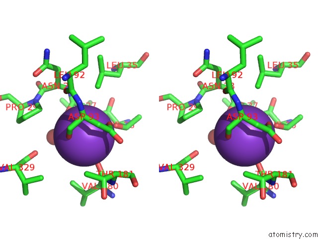
Potassium binding site 2 out of 8 in 2xdr
Go back to
Potassium binding site 2 out
of 8 in the Crystallographic Structure of Betaine Aldehyde Dehydrogenase Mutant E252A From Pseudomonas Aeruginosa
Mono view
Stereo pair view
Mono view
Stereo pair view
A full contact list of Potassium with other atoms in the K binding
site number 2 of Crystallographic Structure of Betaine Aldehyde Dehydrogenase Mutant E252A From Pseudomonas Aeruginosa within 5.0Å range:
|
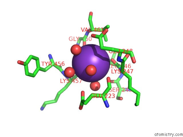
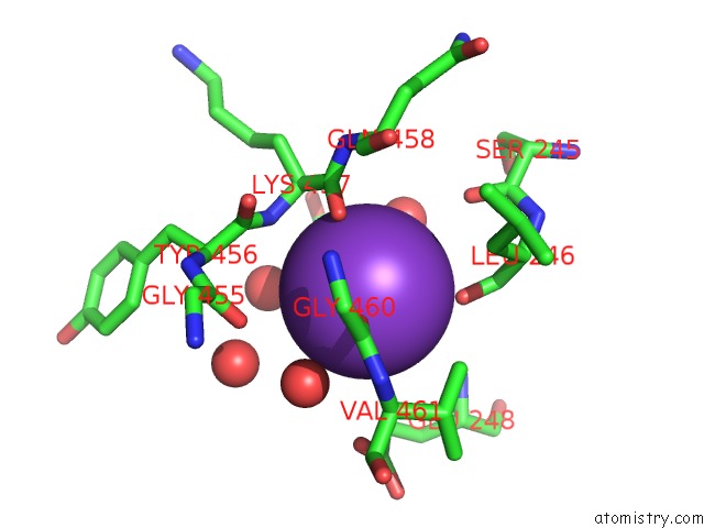
Potassium binding site 3 out of 8 in 2xdr
Go back to
Potassium binding site 3 out
of 8 in the Crystallographic Structure of Betaine Aldehyde Dehydrogenase Mutant E252A From Pseudomonas Aeruginosa
Mono view
Stereo pair view
Mono view
Stereo pair view
A full contact list of Potassium with other atoms in the K binding
site number 3 of Crystallographic Structure of Betaine Aldehyde Dehydrogenase Mutant E252A From Pseudomonas Aeruginosa within 5.0Å range:
|
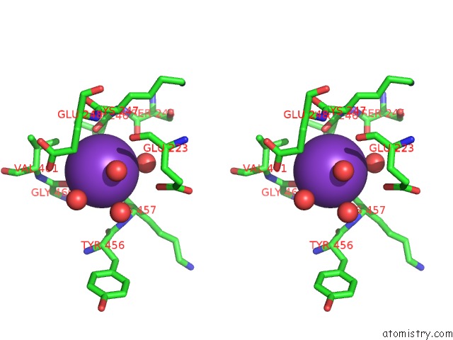
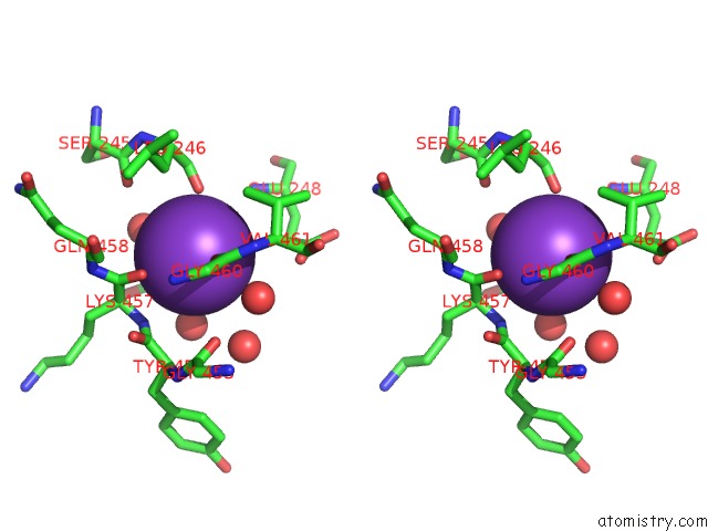
Potassium binding site 4 out of 8 in 2xdr
Go back to
Potassium binding site 4 out
of 8 in the Crystallographic Structure of Betaine Aldehyde Dehydrogenase Mutant E252A From Pseudomonas Aeruginosa
Mono view
Stereo pair view
Mono view
Stereo pair view
A full contact list of Potassium with other atoms in the K binding
site number 4 of Crystallographic Structure of Betaine Aldehyde Dehydrogenase Mutant E252A From Pseudomonas Aeruginosa within 5.0Å range:
|
Potassium binding site 5 out of 8 in 2xdr
Go back to
Potassium binding site 5 out
of 8 in the Crystallographic Structure of Betaine Aldehyde Dehydrogenase Mutant E252A From Pseudomonas Aeruginosa
Mono view
Stereo pair view
Mono view
Stereo pair view
A full contact list of Potassium with other atoms in the K binding
site number 5 of Crystallographic Structure of Betaine Aldehyde Dehydrogenase Mutant E252A From Pseudomonas Aeruginosa within 5.0Å range:
|
Potassium binding site 6 out of 8 in 2xdr
Go back to
Potassium binding site 6 out
of 8 in the Crystallographic Structure of Betaine Aldehyde Dehydrogenase Mutant E252A From Pseudomonas Aeruginosa
Mono view
Stereo pair view
Mono view
Stereo pair view
A full contact list of Potassium with other atoms in the K binding
site number 6 of Crystallographic Structure of Betaine Aldehyde Dehydrogenase Mutant E252A From Pseudomonas Aeruginosa within 5.0Å range:
|
Potassium binding site 7 out of 8 in 2xdr
Go back to
Potassium binding site 7 out
of 8 in the Crystallographic Structure of Betaine Aldehyde Dehydrogenase Mutant E252A From Pseudomonas Aeruginosa
Mono view
Stereo pair view
Mono view
Stereo pair view
A full contact list of Potassium with other atoms in the K binding
site number 7 of Crystallographic Structure of Betaine Aldehyde Dehydrogenase Mutant E252A From Pseudomonas Aeruginosa within 5.0Å range:
|
Potassium binding site 8 out of 8 in 2xdr
Go back to
Potassium binding site 8 out
of 8 in the Crystallographic Structure of Betaine Aldehyde Dehydrogenase Mutant E252A From Pseudomonas Aeruginosa
Mono view
Stereo pair view
Mono view
Stereo pair view
A full contact list of Potassium with other atoms in the K binding
site number 8 of Crystallographic Structure of Betaine Aldehyde Dehydrogenase Mutant E252A From Pseudomonas Aeruginosa within 5.0Å range:
|
Reference:
A.G.Diaz-Sanchez,
L.Gonzalez-Segura,
E.Rudino-Pinera,
A.Lira-Rocha,
R.A.Munoz-Clares.
A Novel Cysteine-Nadph Covalent Adduct in Pseudomonas Aeruginosa Betaine Aldehyde Dehydrogenase Suggests Important Roles For the Reduced Nucleotide in the Reaction Mechanism To Be Published.
Page generated: Mon Aug 12 07:37:24 2024
Last articles
Zn in 9MJ5Zn in 9HNW
Zn in 9G0L
Zn in 9FNE
Zn in 9DZN
Zn in 9E0I
Zn in 9D32
Zn in 9DAK
Zn in 8ZXC
Zn in 8ZUF