Potassium »
PDB 2w0f-2xo0 »
2wme »
Potassium in PDB 2wme: Crystallographic Structure of Betaine Aldehyde Dehydrogenase From Pseudomonas Aeruginosa
Enzymatic activity of Crystallographic Structure of Betaine Aldehyde Dehydrogenase From Pseudomonas Aeruginosa
All present enzymatic activity of Crystallographic Structure of Betaine Aldehyde Dehydrogenase From Pseudomonas Aeruginosa:
1.2.1.8;
1.2.1.8;
Protein crystallography data
The structure of Crystallographic Structure of Betaine Aldehyde Dehydrogenase From Pseudomonas Aeruginosa, PDB code: 2wme
was solved by
L.Gonzalez-Segura,
E.Rudino-Pinera,
R.A.Munoz-Clares,
E.Horjales,
with X-Ray Crystallography technique. A brief refinement statistics is given in the table below:
| Resolution Low / High (Å) | 42.68 / 2.10 |
| Space group | C 1 2 1 |
| Cell size a, b, c (Å), α, β, γ (°) | 334.947, 133.011, 101.814, 90.00, 94.94, 90.00 |
| R / Rfree (%) | 16.598 / 21.123 |
Potassium Binding Sites:
Pages:
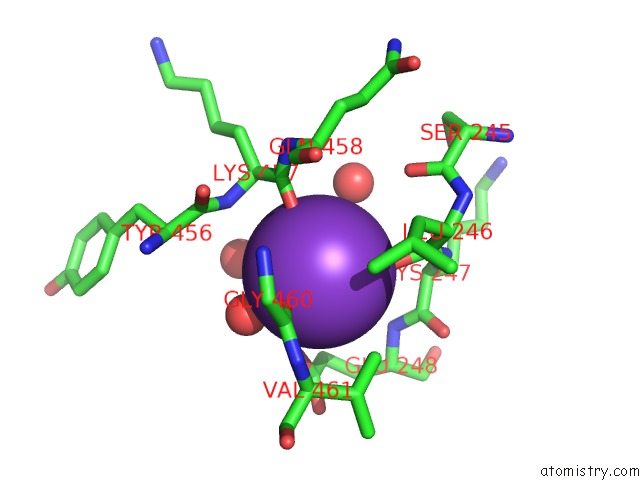
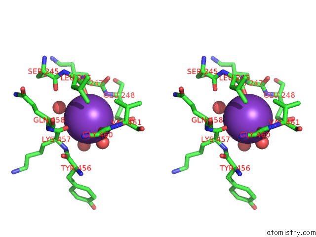
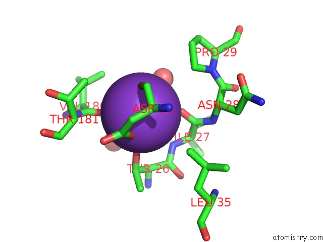
>>> Page 1 <<< Page 2, Binding sites: 11 - 16;Binding sites:
The binding sites of Potassium atom in the Crystallographic Structure of Betaine Aldehyde Dehydrogenase From Pseudomonas Aeruginosa (pdb code 2wme). This binding sites where shown within 5.0 Angstroms radius around Potassium atom.In total 16 binding sites of Potassium where determined in the Crystallographic Structure of Betaine Aldehyde Dehydrogenase From Pseudomonas Aeruginosa, PDB code: 2wme:
Jump to Potassium binding site number: 1; 2; 3; 4; 5; 6; 7; 8; 9; 10;
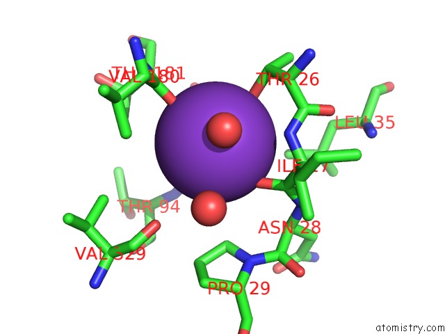
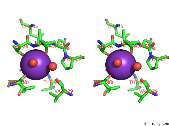
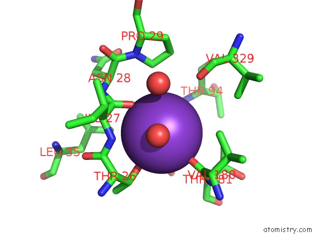
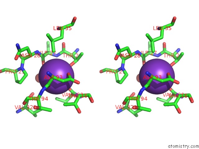
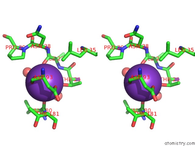
Potassium binding site 1 out of 16 in 2wme
Go back to
Potassium binding site 1 out
of 16 in the Crystallographic Structure of Betaine Aldehyde Dehydrogenase From Pseudomonas Aeruginosa
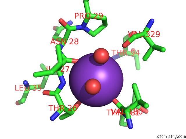
Mono view
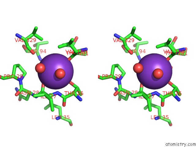
Stereo pair view
Mono view
Stereo pair view
A full contact list of Potassium with other atoms in the K binding
site number 1 of Crystallographic Structure of Betaine Aldehyde Dehydrogenase From Pseudomonas Aeruginosa within 5.0Å range:
|
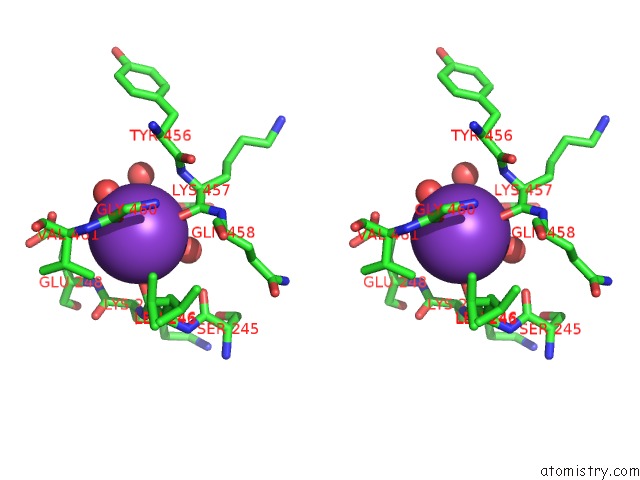
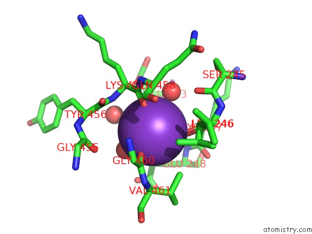
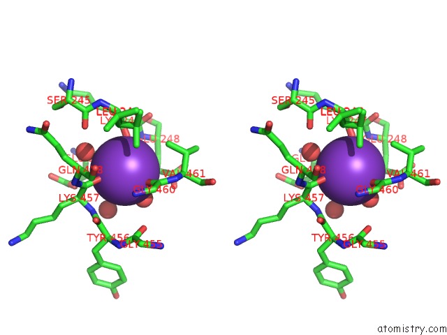
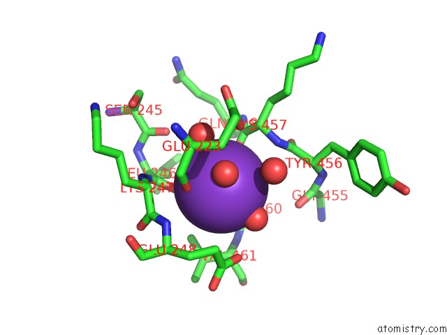
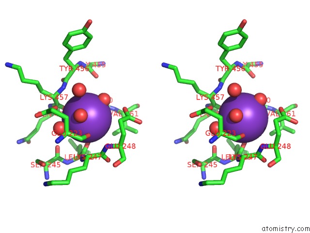
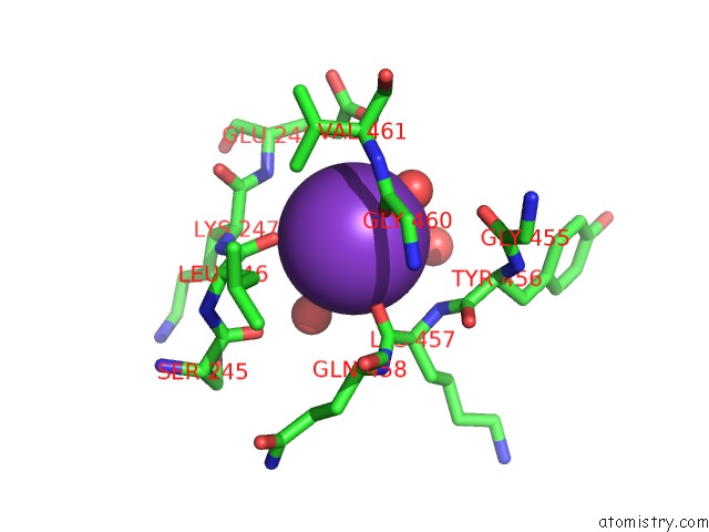
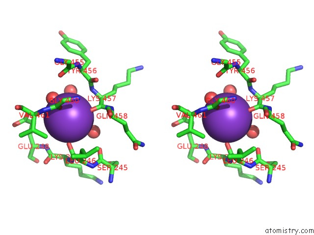
Potassium binding site 2 out of 16 in 2wme
Go back to
Potassium binding site 2 out
of 16 in the Crystallographic Structure of Betaine Aldehyde Dehydrogenase From Pseudomonas Aeruginosa
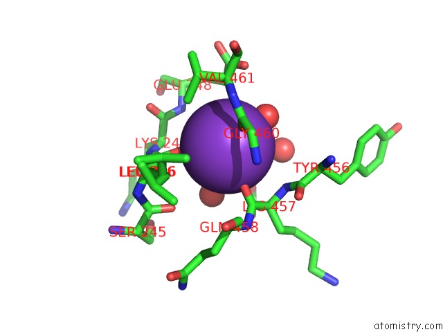
Mono view
Stereo pair view
Mono view
Stereo pair view
A full contact list of Potassium with other atoms in the K binding
site number 2 of Crystallographic Structure of Betaine Aldehyde Dehydrogenase From Pseudomonas Aeruginosa within 5.0Å range:
|
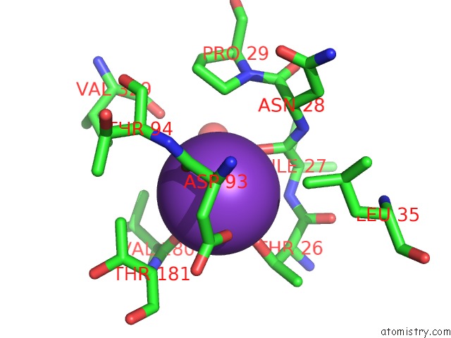
Potassium binding site 3 out of 16 in 2wme
Go back to
Potassium binding site 3 out
of 16 in the Crystallographic Structure of Betaine Aldehyde Dehydrogenase From Pseudomonas Aeruginosa
Mono view
Stereo pair view
Mono view
Stereo pair view
A full contact list of Potassium with other atoms in the K binding
site number 3 of Crystallographic Structure of Betaine Aldehyde Dehydrogenase From Pseudomonas Aeruginosa within 5.0Å range:
|
Potassium binding site 4 out of 16 in 2wme
Go back to
Potassium binding site 4 out
of 16 in the Crystallographic Structure of Betaine Aldehyde Dehydrogenase From Pseudomonas Aeruginosa
Mono view
Stereo pair view
Mono view
Stereo pair view
A full contact list of Potassium with other atoms in the K binding
site number 4 of Crystallographic Structure of Betaine Aldehyde Dehydrogenase From Pseudomonas Aeruginosa within 5.0Å range:
|
Potassium binding site 5 out of 16 in 2wme
Go back to
Potassium binding site 5 out
of 16 in the Crystallographic Structure of Betaine Aldehyde Dehydrogenase From Pseudomonas Aeruginosa
Mono view

Stereo pair view
Mono view
Stereo pair view
A full contact list of Potassium with other atoms in the K binding
site number 5 of Crystallographic Structure of Betaine Aldehyde Dehydrogenase From Pseudomonas Aeruginosa within 5.0Å range:
|
Potassium binding site 6 out of 16 in 2wme
Go back to
Potassium binding site 6 out
of 16 in the Crystallographic Structure of Betaine Aldehyde Dehydrogenase From Pseudomonas Aeruginosa
Mono view
Stereo pair view
Mono view
Stereo pair view
A full contact list of Potassium with other atoms in the K binding
site number 6 of Crystallographic Structure of Betaine Aldehyde Dehydrogenase From Pseudomonas Aeruginosa within 5.0Å range:
|
Potassium binding site 7 out of 16 in 2wme
Go back to
Potassium binding site 7 out
of 16 in the Crystallographic Structure of Betaine Aldehyde Dehydrogenase From Pseudomonas Aeruginosa
Mono view
Stereo pair view
Mono view
Stereo pair view
A full contact list of Potassium with other atoms in the K binding
site number 7 of Crystallographic Structure of Betaine Aldehyde Dehydrogenase From Pseudomonas Aeruginosa within 5.0Å range:
|
Potassium binding site 8 out of 16 in 2wme
Go back to
Potassium binding site 8 out
of 16 in the Crystallographic Structure of Betaine Aldehyde Dehydrogenase From Pseudomonas Aeruginosa
Mono view
Stereo pair view
Mono view
Stereo pair view
A full contact list of Potassium with other atoms in the K binding
site number 8 of Crystallographic Structure of Betaine Aldehyde Dehydrogenase From Pseudomonas Aeruginosa within 5.0Å range:
|
Potassium binding site 9 out of 16 in 2wme
Go back to
Potassium binding site 9 out
of 16 in the Crystallographic Structure of Betaine Aldehyde Dehydrogenase From Pseudomonas Aeruginosa
Mono view
Stereo pair view
Mono view
Stereo pair view
A full contact list of Potassium with other atoms in the K binding
site number 9 of Crystallographic Structure of Betaine Aldehyde Dehydrogenase From Pseudomonas Aeruginosa within 5.0Å range:
|
Potassium binding site 10 out of 16 in 2wme
Go back to
Potassium binding site 10 out
of 16 in the Crystallographic Structure of Betaine Aldehyde Dehydrogenase From Pseudomonas Aeruginosa
Mono view
Stereo pair view
Mono view
Stereo pair view
A full contact list of Potassium with other atoms in the K binding
site number 10 of Crystallographic Structure of Betaine Aldehyde Dehydrogenase From Pseudomonas Aeruginosa within 5.0Å range:
|
Reference:
L.Gonzalez-Segura,
E.Rudino-Pinera,
R.A.Munoz-Clares,
E.Horjales.
The Crystal Structure of A Ternary Complex of Betaine Aldehyde Dehydrogenase From Pseudomonas Aeruginosa Provides New Insight Into the Reaction Mechanism and Shows A Novel Binding Mode of the 2'- Phosphate of Nadp(+) and A Novel Cation Binding Site. J.Mol.Biol. V. 385 542 2009.
ISSN: ISSN 0022-2836
PubMed: 19013472
DOI: 10.1016/J.JMB.2008.10.082
Page generated: Sat Aug 9 04:19:36 2025
ISSN: ISSN 0022-2836
PubMed: 19013472
DOI: 10.1016/J.JMB.2008.10.082
Last articles
Mg in 1HPMMg in 1HN1
Mg in 1HC8
Mg in 1HMV
Mg in 1HI0
Mg in 1HJK
Mg in 1HK7
Mg in 1HI8
Mg in 1HJ6
Mg in 1HBN